. 2006 Dec;147(12):5817-25.
doi: 10.1210/en.2006-0787. Epub 2006 Sep 7.

Postnatal development of kisspeptin neurons in mouse hypothalamus; sexual dimorphism and projections to gonadotropin-releasing hormone neurons

Affiliations

Postnatal development of kisspeptin neurons in mouse hypothalamus; sexual dimorphism and projections to gonadotropin-releasing hormone neurons

Jenny Clarkson et al. Endocrinology. 2006 Dec.

Abstract

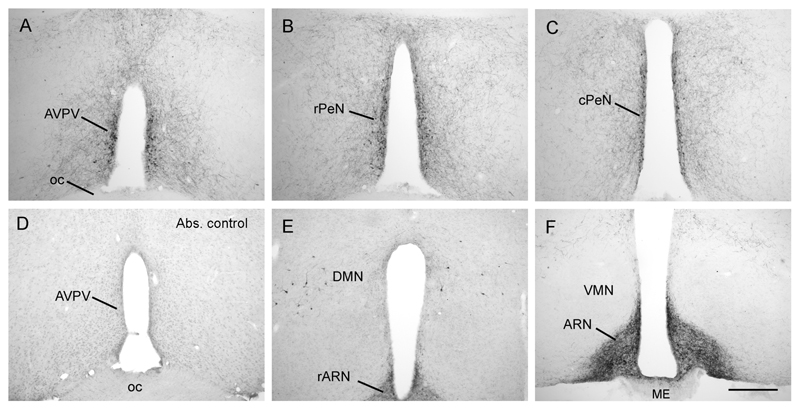
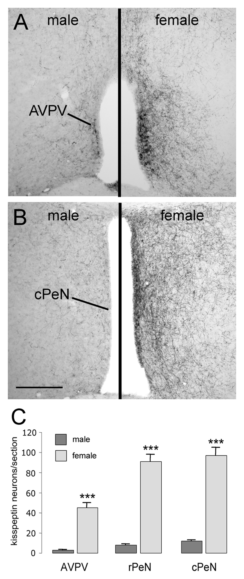
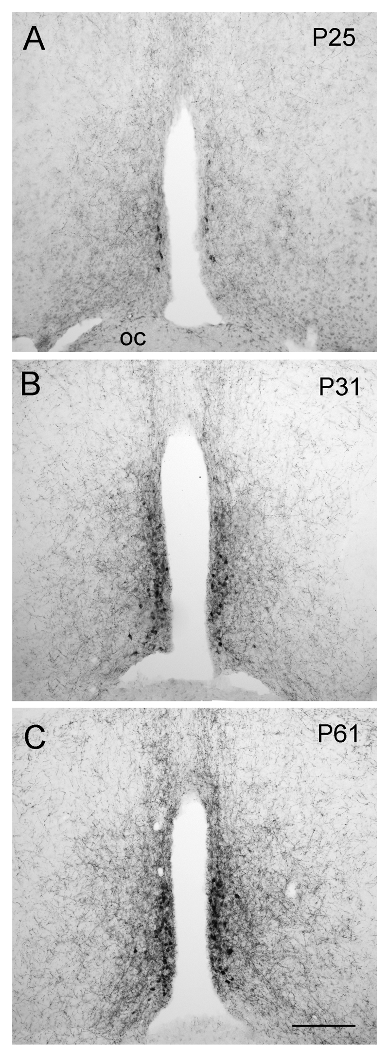
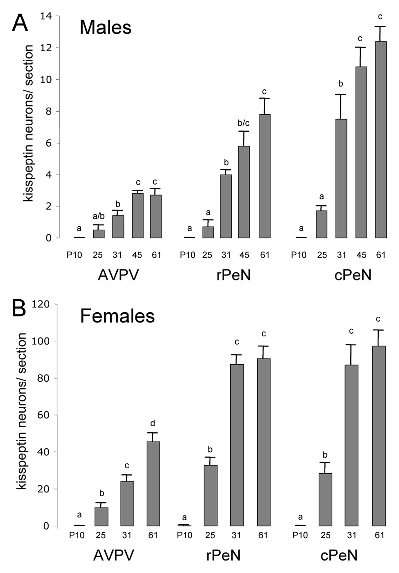
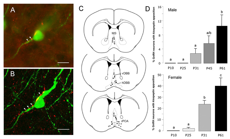
The neuropeptide kisspeptin has recently been implicated as having a critical role in the activation of the GnRH neurons to bring about puberty. We examined here the postnatal development of kisspeptin neuronal populations and their projections to GnRH neurons in the mouse. Three populations of kisspeptin neurons located in the 1) anteroventral periventricular nucleus (AVPV) and the preoptic periventricular nucleus (PeN), 2) dorsomedial hypothalamus, and 3) arcuate nucleus were identified using an antisera raised against mouse kisspeptin-10. A marked 10-fold (P<0.01), female-dominant sex difference in the numbers of kisspeptin neurons existed in the AVPV/PeN but not elsewhere. Kisspeptin neurons in the AVPV/PeN of both sexes displayed a similar pattern of postnatal development with no cells detected at postnatal day (P) 10, followed by increases from P25 to reach adult levels by puberty onset (P<0.01; P31 females and P45 males). This pattern was not found in the dorsomedial hypothalamus or arcuate nucleus. Dual immunofluorescence experiments demonstrated close appositions between kisspeptin fibers and GnRH neuron cell bodies that were first apparent at P25 and increased across postnatal development in both sexes. These studies demonstrate kisspeptin peptide expression in the mouse hypothalamus and reveal the postnatal development of a sexually dimorphic continuum of kisspeptin neurons within the AVPV and PeN. This periventricular population of kisspeptin neurons reaches adult-like proportions at the time of puberty onset and is the likely source of the kisspeptin inputs to GnRH neurons.


Conflict of interest statement

Author Disclosure Statement

The authors have nothing to declare.

Figures

Figure 1
Figure 1. Distribution of kisspeptin immunoreactivity in the mouse hypothalamus.
Kisspeptin cell bodies were found in a periventricular continuum beginning in the anteroventral periventricular nucleus (AVPV; A) and extending caudally through into the preoptic periventricular nucleus (PeN), divided here into the rostral (rPeN; B) and caudal (cPeN; C) halves for analyses. There was a complete absence of labelling in tissue that was incubated with kisspeptin-10 antisera that was preadsorbed with mouse kisspeptin-10 peptide (D). Kisspeptin cell bodies were also present scattered within the dorsomedial nucleus (DMN; E), while dense fiber staining filled the arcuate nucleus (ARN; F) with cell bodies only occasionally discernable. ME, median eminence; oc, optic chiasm; VMN, ventromedial nucleus. Scale bar in F is 300μm.
Figure 2
Figure 2. Distribution of kisspeptin immunoreactive fibers in the mouse hypothalamus.
A low-power view of kisspeptin fibers within the rostral preoptic area where the largest density of GnRH neuron cell bodies are found. B same region but following staining with kisspeptin antiserum adsorbed with kisspeptin-10. C Higher-power view of kisspeptin fiber plexus within the arcuate nucleus (ARN). Note the cell body profile at the ventral edge of the ARN and lack of staining in the external zone of the median eminence (ME). D same region but following staining with kisspeptin antiserum adsorbed with kisspeptin-10. Kisspeptin fiber staining was also found in the supraoptic (E) and paraventricular (F) nuclei. G and H show high-power images of kisspeptin-immunoreactive fiber and cell bodies in the preoptic periventricular nucleus in postnatal day 61 (P61) and P25 female mice, respectively. Abbreviations = MnPO, median preoptic nucleus; OVLT, organum vasculosum of the lamina terminalis; IIIV, third ventricle; ME, median eminence; OC, optic chiasm; SON, supraoptic nucleus; PVN, paraventricular nucleus. Scale bars represent 240μm (A,B,E,F) and 120μm (C,D,G,H).
Figure 3
Figure 3. Sex differences in kisspeptin immunoreactivity in the rostral hypothalamus.
Panels A and B show kisspeptin immunoreactivity in the adult male (left panel) and female (right panel) anteroventral periventricular nucleus (AVPV; A) and caudal preoptic periventricular nucleus (cPeN; B). Quantitative analyses (mean +SEM) of numbers of kisspeptin-immunoreactive cell bodies in the AVPV, rostral and caudal halves of the PeN are given in C. ** p<0.01, *** p<0.001; male (n=6), female (n=4). Scale bar in B is 300μm.
Figure 4
Figure 4. Developmental increase in kisspeptin staining in the periventricular nucleus.
Kisspeptin-immunoreactivity in postnatal day 25 (P25), P31 and adult (P61) female mice within the rostral hypothalamus. Scale bar in C is 150μm.
Figure 5
Figure 5. Quantitative analysis of kisspeptin-immunoreactive cell bodies in the developing periventricular nuclei.
Data are shown for the anteroventral periventricular (AVPV) and preoptic periventricular nucleus (PeN) divided into rostral (rPeN) and caudal (cPeN) halves. The mean (+SEM) number of immunoreactive cells detected per section at the three levels and at the indicated postnatal days (P) is given for males (A) and females (B). Bars labeled with different letters are significantly different from each other at either p<0.05 or p<0.01 (see text). N= 4-8 for each sex and age group.
Figure 6
Figure 6. Postnatal development of kisspeptin fiber projections to GnRH neurons.
A. Confocal stack of 75 images showing a single GnRH neuron (green) with kisspeptin (red) fibers surrounding and opposed to it. Single 370nm-thick optical sections through the three regions indicated by a, b, c of the GnRH neuron are given below to demonstrate the close apposition between kisspeptin fibers and GnRH neuron elements. Scale bar represents 10μm. B depicts the levels at which GnRH neurons were analysed including the medial septum (MS), vertical limb of the diagonal band of Broca (vDBB), horizontal limb (hDBB) and the rostral preoptic area (rPOA). The distribution of GnRH neurons exhibiting kisspeptin fiber appositions is shown for an adult female mouse (open circles represent GnRH neurons with no close apposition; filled circles are GnRH neurons with kisspeptin appositions). C depicts the mean (+SEM) percentage of GnRH neurons with kisspeptin fiber appositions in males (top) and females (bottom) at different postnatal (P) ages. Bars labelled with different letters are significantly different from each other at p<0.05.

References

    1. Terasawa E, Fernandez DL. Neurobiological mechanisms of the onset of puberty in primates. Endocr Rev. 2001;22:111–151. - PubMed
    1. Ojeda SR, Skinner MK. Puberty in the rat. In: Neill JD, editor. Knobil and Neill's Physiology of Reproduction. 3rd ed. San Diego: Academic Press; 2006. pp. 2061–2126.
    1. Plant TM, Witchel SF. Puberty in nonhuman primates and humans. In: Neill JD, editor. Knobil and Neill's Physiology of Reproduction. 3rd ed. San Diego: Academic Press; 2006. pp. 2177–2230.
    1. Kotani M, Detheux M, Vandenbogaerde A, Communi D, Vanderwinden JM, Le Poul E, Brezillon S, Tyldesley R, Suarez-Huerta N, Vandeput F, Blanpain C, et al. The metastasis suppressor gene KiSS-1 encodes kisspeptins, the natural ligands of the orphan G protein-coupled receptor GPR54. J Biol Chem. 2001;276:34631–34636. - PubMed
    1. Muir AI, Chamberlain L, Elshourbagy NA, Michalovich D, Moore DJ, Calamari A, Szekeres PG, Sarau HM, Chambers JK, Murdock P, Steplewski K, et al. AXOR12, a novel human G protein-coupled receptor, activated by the peptide KiSS-1. J Biol Chem. 2001;276:28969–28975. - PubMed

Publication types

MeSH terms