. 2006 Sep 19;103(38):13932-7.
doi: 10.1073/pnas.0606411103. Epub 2006 Sep 7.

Disulfide isomerization switches tissue factor from coagulation to cell signaling

Affiliations

Disulfide isomerization switches tissue factor from coagulation to cell signaling

Jasimuddin Ahamed et al. Proc Natl Acad Sci U S A. .

Abstract

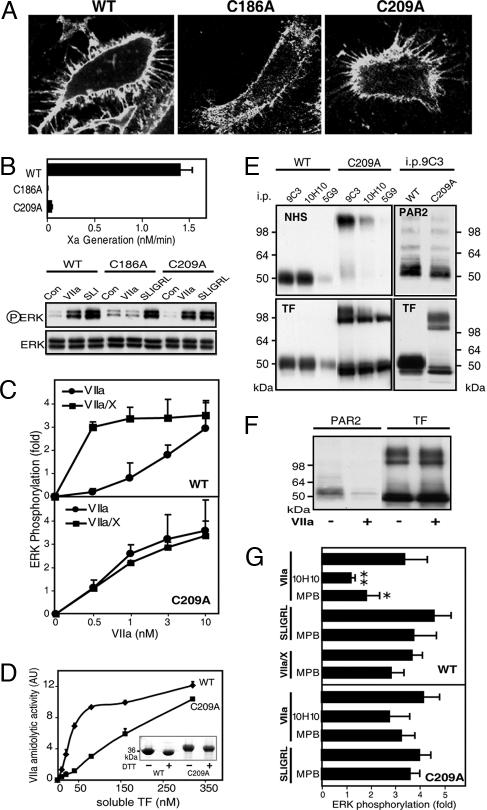
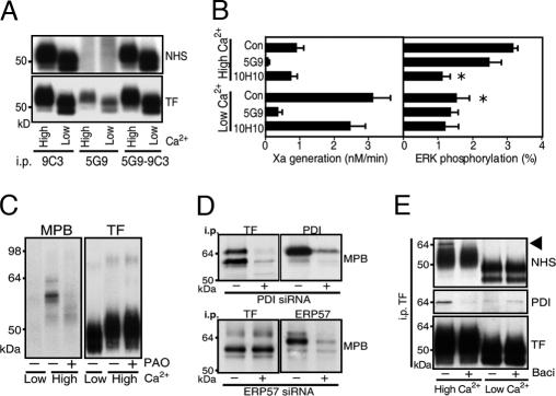
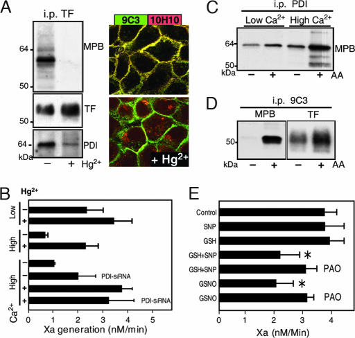
Cell-surface tissue factor (TF) binds the serine protease factor VIIa to activate coagulation or, alternatively, to trigger signaling through the G protein-coupled, protease-activated receptor 2 (PAR2) relevant to inflammation and angiogenesis. Here we demonstrate that TF.VIIa-mediated coagulation and cell signaling involve distinct cellular pools of TF. The surface-accessible, extracellular Cys186-Cys209 disulfide bond of TF is critical for coagulation, and protein disulfide isomerase (PDI) disables coagulation by targeting this disulfide. A TF mutant (TF C209A) with an unpaired Cys186 retains TF.VIIa signaling activity, and it has reduced affinity for VIIa, a characteristic of signaling TF on cells with constitutive TF expression. We further show that PDI suppresses TF coagulant activity in a nitric oxide-dependent pathway, linking the regulation of TF thrombogenicity to oxidative stress in the vasculature. Furthermore, a unique monoclonal antibody recognizes only the noncoagulant, cryptic conformation of TF. This antibody inhibits formation of the TF.PAR2 complex and TF.VIIa signaling, but it does not prevent coagulation activation. These experiments delineate an upstream regulatory mechanism that controls TF function, and they provide initial evidence that TF.VIIa signaling can be specifically inhibited with minimal effects on coagulation.


Conflict of interest statement

Conflict of interest statement: No conflicts declared.

Figures

Fig. 1.
Fig. 1.
TF·VIIa signaling and coagulation initiation are mediated by distinct cell-surface pools of TF. (A) TF coagulant activity measured as Xa generation and TF expression quantified from Western blots during growth arrest of HaCaT cells. Results are expressed as the mean ± SD (n = 3). (Insets) Representative Western blots for actin or TF in cell lysates. (B) Detection of cell-surface TF with FITC-conjugated mAb 9C3 and Texas red-conjugated mAb 5G9 or 10H10 by confocal microscopy. (C) mAb 5G9 but not mAb 10H10 inhibits TF coagulant activity equally during growth arrest. (D) Immobilized mAb 5G9 but not mAb 10H10 immunodepletes coagulation activity from preparations of phospholipid-reconstituted TF. (E) TF·VIIa signaling is down-regulated during growth arrest. (Insets) Representative Western blots for phosphorylated and nonphosphorylated ERK after 10 min of stimulation. (F) PAR2 agonist SLIGRL and thrombin responses are not down-regulated during growth arrest. Pretreatment of cells at day 1 with inhibitory antibodies to PAR1 or PAR2 shows PAR2 dependence of TF·VIIa signaling. (G) mAb 10H10 specifically inhibits TF·VIIa signaling. Different affinity for VIIa distinguishes TF that mediates TF·VIIa or ternary TF·VIIa·Xa complex signaling. At 1 nM VIIa, cells display only Xa-dependent TF·VIIa·Xa signaling that is inhibited by the specific Xa inhibitor nematode anticoagulant protein 5 (NAP5) or mAb 5G9. At 10 nM VIIa, direct signaling of TF·VIIa occurs that is not inhibited by NAP5 or mAb 5G9 but is blocked by mAb 10H10. ERK phosphorylation at 10 min was quantified (mean ± SD; n > 3).
Fig. 2.
Fig. 2.
The TF Cys186–Cys209 disulfide controls TF signaling specificity. (A) Cell-surface expression shown by FITC-labeled mAb 9C3 staining of wild-type (WT), C186A, or C209A TF in HUVECs cotransduced with PAR2. (B) Mutation of Cys186 or Cys209 reduces coagulation, but only C209A TF retains TF·VIIa signaling activity. Control (Con) experiments (not shown) revealed no VIIa signaling in cells that were transduced only with PAR2 or vector control, excluding that adenovirus transduction induced the up-regulation of endogenous TF under these conditions. (C) Dose–response of VIIa signaling with and without 100 nM X in HUVECs expressing C209A or WT TF. ERK phosphorylation was quantified at 10 min (mean ± SD; n > 4). (D) Recombinant soluble C209A TF enhances catalytic activity of VIIa (40 nM) with reduced affinity relative to WT TF (mean ± SD; n = 3). AU, arbitrary units. (Inset) Gel of homogeneous preparations of monomeric soluble TF; the expression tag was not cleaved from C209A TF, yielding a higher molecular mass. (E) Cell-surface expression of WT and C209A TF determined by NHS surface biotinylation. mAb 5G9 epitope loss after surface NHS modification was used to demonstrate relative abundance of intra- and extracellular pools. The majority of cell-surface C209A TF was present as an SDS-stable homodimer with a consistently observed pool of C209A TF monomer in PAR2-transduced cells. PAR2 coimmunoprecipitated (i.p.) with WT or C209A TF in an SDS-labile complex. (F) PAR2 detection in mAb 9C3 immunoprecipitates of C209A TF is sensitive to pretreatment of cells with VIIa. (G) Inhibition of WT TF·VIIa signaling by thiol blockade. TF- and PAR2-transduced HUVECs were pretreated with 100 μM MPB or 50 μg/ml mAb 10H10 for 15 min before stimulation with 10 nM VIIa, 0.5 nM VIIa, and 100 nM X, or SLIGRL (mean ± SD; n = 3).
Fig. 3.
Fig. 3.
Extracellular PDI is associated with TF. (A) A 24-h elevation of extracellular Ca2+ yields similar TF cell surface expression relative to HaCaT cells in low Ca2+. TF was immunoprecipitated (i.p.) from NHS surface-biotinylated cells with mAb 9C3 or 5G9. mAb 5G9 does not bind biotinylated TF that is recovered in subsequent mAb 9C3 pulldown. (B) Low coagulant activity of TF in high-Ca2+ cells is associated with TF·VIIa signaling that is inhibited by mAb 10H10. ∗, Different from high-Ca2+ control (P < 0.01, t test; mean ± SD; n > 4). (C) MPB labeling of proteins coprecipitating with TF mAb 9C3 pulldown is inhibited by blockade of vicinal thiols by 2 μM phenylarsine oxide (PAO). (D) PDI but not ERP57 knockdown with siRNA prevents MPB-labeled bands in TF immunoprecipitates. (E) NHS surface-biotinylated PDI is specifically associated with high-Ca2+ cells. The PDI inhibitor bacitracin (3 mM) also dissociates PDI from TF in mAb 9C3 immunoprecipitates, but it has no effect on low-Ca2+ cells.
Fig. 4.
Fig. 4.
NO-dependent suppression of TF coagulant activity by PDI. (A) A 2-min exposure to 100 μM Hg2+ dissociates MPB-labeled PDI from anti-TF mAb 9C3 immunoprecipitates (i.p.) and abolishes mAb 10H10 staining of TF. (B) Hg2+ treatment induces coagulation specifically on high-Ca2+ cells. Knockdown of PDI in high-Ca2+ cells increases TF coagulant activity without changing maximal function after Hg2+ treatment. (C) The biotin-switch method after thiol blockade with 1 mM N-ethylmaleimide detects increased labeling of PDI after NO release by ascorbic acid (AA) specifically in PDI immunoprecipitates from high-Ca2+ cells. (D) S-nitrosylation of TF detected by the biotin-switch method after thiol blockade with 1 mM iodoacetamide before MPB-labeling with or without ascorbic acid. (E) Hg2+-induced activation of TF coagulant activity is reversible by NO-dependent PDI pathways. Cells washed after brief 100 μM Hg2+ exposure were incubated in Hepes buffer, pH 7.4/1.5 mM Ca2+ in the presence of 1 mM SNP, 1 mM reduced glutathione (GSH), or 1 mM S-nitrosoglutathione (GSNO) with or without 10 μM PAO for 10 min before the Xa generation assay. *, Different from control (P < 0.05, t test; mean ± SD; n = 3).
Fig. 5.
Fig. 5.
TF·PAR2 complex formation is required for TF·VIIa signaling. (A) mAb 5G9 immunoprecipitation (i.p.) of PAR2 from high-Ca2+ cells is abolished by Hg2+ pretreatment. (B) mAb 10H10 but not mAb 5G9 perturbs the TF·PAR2 complex. HaCaT cells were pretreated in serum-free medium for 15 min with the indicated antibody, and TF was detected in the PAR2 immunoprecipitate. Loading controls were not feasible due to background because no PAR2-suitable antibody from a different species was available for Western blotting. (C) mAb 10H10 does not immunoprecipitate PAR2 from HaCaT cells. (D) mAb 10H10 does not immunoprecipitate a complex containing PDI. MPB-labeled cells were immunoprecipitated with mAb 9C3 or mAb 10H10 and probed for PDI, TF, or thiol biotinylation with MPB. (E) Dissociation of PDI from TF by bacitracin is reversible. (F) Bacitracin reversibly blocks TF·VIIa signaling. Wash out indicates that cells preincubated for 10 min with bacitracin were washed and equilibrated in serum-free medium for 10 min before stimulation with 10 nM VIIa. *, P < 0.01 relative to control (t test; mean ± SD; n > 4).

Comment in

References

    1. Norledge B, Petrovan RJ, Ruf W, Olson A. Proteins. 2003;53:640–648. - PubMed
    1. Mackman N. Arterioscler Thromb Vasc Biol. 2004;24:1015–1022. - PubMed
    1. Camerer E, Huang W, Coughlin SR. Proc Natl Acad Sci USA. 2000;97:5255–5260. - PMC - PubMed
    1. Riewald M, Ruf W. Proc Natl Acad Sci USA. 2001;98:7742–7747. - PMC - PubMed
    1. Riewald M, Ruf W. Crit Care. 2003;7:123–129. - PMC - PubMed

Publication types

LinkOut - more resources