Review
. 2006 Sep;70(3):755-88.
doi: 10.1128/MMBR.00008-06.

Mapping the pathways to staphylococcal pathogenesis by comparative secretomics

Affiliations
Review

Mapping the pathways to staphylococcal pathogenesis by comparative secretomics

M J J B Sibbald et al. Microbiol Mol Biol Rev. 2006 Sep.

Abstract

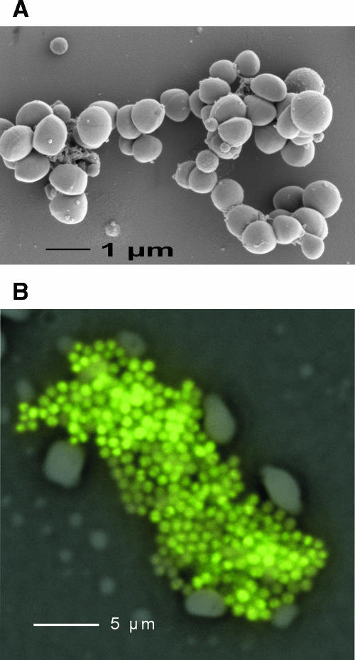
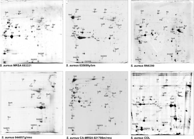
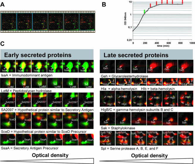
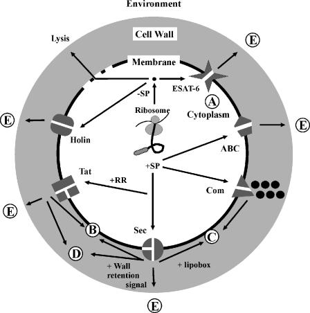
The gram-positive bacterium Staphylococcus aureus is a frequent component of the human microbial flora that can turn into a dangerous pathogen. As such, this organism is capable of infecting almost every tissue and organ system in the human body. It does so by actively exporting a variety of virulence factors to the cell surface and extracellular milieu. Upon reaching their respective destinations, these virulence factors have pivotal roles in the colonization and subversion of the human host. It is therefore of major importance to obtain a clear understanding of the protein transport pathways that are active in S. aureus. The present review aims to provide a state-of-the-art roadmap of staphylococcal secretomes, which include both protein transport pathways and the extracytoplasmic proteins of these organisms. Specifically, an overview is presented of the exported virulence factors, pathways for protein transport, signals for cellular protein retention or secretion, and the exoproteomes of different S. aureus isolates. The focus is on S. aureus, but comparisons with Staphylococcus epidermidis and other gram-positive bacteria, such as Bacillus subtilis, are included where appropriate. Importantly, the results of genomic and proteomic studies on S. aureus secretomes are integrated through a comparative "secretomics" approach, resulting in the first definition of the core and variant secretomes of this bacterium. While the core secretome seems to be largely employed for general housekeeping functions which are necessary to thrive in particular niches provided by the human host, the variant secretome seems to contain the "gadgets" that S. aureus needs to conquer these well-protected niches.


Figures

FIG. 1.
FIG. 1.
Imaging of S. aureus RN6390. (A) For scanning electron microscopy, a drop of washed culture of bacteria was fixated for 30 min with 2% glutaraldehyde in 0.1 M cacodylate buffer, pH 7.38. Next, the fixated bacteria were placed on a piece (1 cm2) of cleaved 0.1% poly-l-lysine-coated mica sheet and washed in 0.1 M cacodylate buffer. This specimen was dehydrated in an ethanol series consisting of 30%, 50%, 70%, 96%, and anhydrous 100% (3×) solutions for 10 min each, critical point dried with CO2, and sputter coated with 2 to 3 nm Au/Pd (Balzers coater). The specimen was fixed on a scanning electron microscope stub holder and observed in a JEOL FE-SEM 6301F microscope. (B) Micrograph of a cluster of S. aureus cells grown in blood culture medium. The cells were fixed with ethanol and hybridized with the fluorescein-labeled peptide nucleic acid (PNA) probe PNA-Stau. The image was generated by merging an epifluorescence image with the negative of a phase-contrast image.
FIG. 2.
FIG. 2.
Extracellular proteomes of different S. aureus strains. Proteins in the growth medium fractions of different staphylococcal isolates, grown in TSB medium (37°C) to an optical density at 540 nm (OD540) of 10, were separated by 2D-PAGE using immobilized pH gradient strips in the pH range of 3 to 10 (Amersham Pharmacia Biotech, Piscataway, N.J.). Each gel was loaded with 350 μg protein extracts and, after electrophoresis, stained with colloidal Coomassie blue. Proteins were identified by matrix-assisted laser desorption ionization-time of flight mass spectrometry. The corresponding protein spots are labeled with protein names according to the S. aureus N315 database or NCBI entries for proteins not present in N315. The S. aureus strains that were used in these experiments are RN6390 and COL and four clinical isolates from the University Medical Center Groningen, named MRSA693331, 035699y/bm, 0440579/rmo, and CA-MRSA021708m/rmo.
FIG. 3.
FIG. 3.
Dynamics of the amount of extracellular proteins during growth of S. aureus RN6390 in TSB medium. (A) Individual dual-channel 2D patterns of extracellular proteins during the different phases of the growth curve for cells grown in TSB medium were assembled into a movie. The protein pattern at an OD540 of 1 (labeled in green) was compared with the protein patterns at higher optical densities (labeled in red). As a consequence of dual channel labeling, spots where the intensities do not differ between the compared gels are yellow, and spots with different intensities are either green or red (15). (B) Growth curve for S. aureus RN6390 grown in TSB medium, as determined by OD540 readings. The sampling points for proteomics analyses are indicated by arrows. (C) Proteomic signatures of selected proteins representing different regulatory groups, as revealed by dual-channel imaging. The amounts of the respective proteins at an OD540 of 1 (spots labeled in green) for cells grown in TSB medium were compared with the relative amounts of these proteins at higher optical densities (spots labeled in red). Proteins were stained with Sypro ruby.
FIG. 4.
FIG. 4.
Staphylococcal pathways to pathogenesis. The figure shows a schematic representation of a staphylococcal cell with potential pathways for protein sorting and secretion. (A) Proteins without signal peptides reside in the cytoplasm. (B) Proteins with one or more transmembrane-spanning domains can be inserted into the membrane via the Sec, Tat, or Com pathway. (C) Lipoproteins are exported via the Sec pathway and are anchored to the membrane after lipid modification. (D) Proteins with cell wall retention signals are exported via the Sec, Tat, or Com pathway and retained in the cell wall via covalent or high-affinity binding to cell wall components. (E) Exported proteins with a signal peptide and without a membrane or cell wall retention signal can be secreted into the extracellular milieu via the various indicated pathways.
FIG. 5.
FIG. 5.
General properties and classification of S. aureus signal peptides. Signal peptide properties are based on SPase cleavage sites and the export pathways by which the preproteins are exported. Predicted signal peptides (144) were divided into the following five distinct classes: secretory (Sec-type) signal peptides, twin-arginine (RR/KR) signal peptides, lipoprotein signal peptides, pseudopilin-like signal peptides, and bacteriocin leader peptides. Most of these signal peptides have a tripartite structure, with a positively charged N domain (N) containing lysine and/or arginine residues (indicated by plus signs), a hydrophobic H domain (H, indicated by a black box), and a C domain (C) that specifies the cleavage site for a specific SPase. Where appropriate, the most frequently occurring amino acid residues at particular positions in the signal peptide or mature protein are indicated. Also, the numbers of signal peptides identified for each class and the respective SPase are indicated.

Similar articles

Cited by

References

    1. Adhikari, R. P., and R. P. Novick. 2005. Subinhibitory cerulenin inhibits staphylococcal exoprotein production by blocking transcription rather than by blocking secretion. Microbiology 151:3059-3069. - PubMed
    1. Alami, M., I. Lüke, S. Deitermann, G. Eisner, H. G. Koch, J. Brunner, and M. Müller. 2003. Differential interactions between a twin-arginine signal peptide and its translocase in Escherichia coli. Mol. Cell 12:937-946. - PubMed
    1. Antelmann, H., H. Tjalsma, B. Voigt, S. Ohlmeier, S. Bron, J. M. van Dijl, and M. Hecker. 2001. A proteomic view on genome-based signal peptide predictions. Genome Res. 11:1484-1502. - PubMed
    1. Avison, M. B., P. M. Bennett, R. A. Howe, and T. R. Walsh. 2002. Preliminary analysis of the genetic basis for vancomycin resistance in Staphylococcus aureus strain Mu50. J. Antimicrob. Chemother. 49:255-260. - PubMed
    1. Baba, T., and O. Schneewind. 1996. Target cell specificity of a bacteriocin molecule: a C-terminal signal directs lysostaphin to the cell wall of Staphylococcus aureus. EMBO J. 15:4789-4797. - PMC - PubMed

MeSH terms

Substances