Review
. 2006 Sep;70(3):789-829.
doi: 10.1128/MMBR.00040-05.

Nuclear ADP-ribosylation reactions in mammalian cells: where are we today and where are we going?

Affiliations
Review

Nuclear ADP-ribosylation reactions in mammalian cells: where are we today and where are we going?

Paul O Hassa et al. Microbiol Mol Biol Rev. 2006 Sep.

Abstract

Since poly-ADP ribose was discovered over 40 years ago, there has been significant progress in research into the biology of mono- and poly-ADP-ribosylation reactions. During the last decade, it became clear that ADP-ribosylation reactions play important roles in a wide range of physiological and pathophysiological processes, including inter- and intracellular signaling, transcriptional regulation, DNA repair pathways and maintenance of genomic stability, telomere dynamics, cell differentiation and proliferation, and necrosis and apoptosis. ADP-ribosylation reactions are phylogenetically ancient and can be classified into four major groups: mono-ADP-ribosylation, poly-ADP-ribosylation, ADP-ribose cyclization, and formation of O-acetyl-ADP-ribose. In the human genome, more than 30 different genes coding for enzymes associated with distinct ADP-ribosylation activities have been identified. This review highlights the recent advances in the rapidly growing field of nuclear mono-ADP-ribosylation and poly-ADP-ribosylation reactions and the distinct ADP-ribosylating enzyme families involved in these processes, including the proposed family of novel poly-ADP-ribose polymerase-like mono-ADP-ribose transferases and the potential mono-ADP-ribosylation activities of the sirtuin family of NAD(+)-dependent histone deacetylases. A special focus is placed on the known roles of distinct mono- and poly-ADP-ribosylation reactions in physiological processes, such as mitosis, cellular differentiation and proliferation, telomere dynamics, and aging, as well as "programmed necrosis" (i.e., high-mobility-group protein B1 release) and apoptosis (i.e., apoptosis-inducing factor shuttling). The proposed molecular mechanisms involved in these processes, such as signaling, chromatin modification (i.e., "histone code"), and remodeling of chromatin structure (i.e., DNA damage response, transcriptional regulation, and insulator function), are described. A potential cross talk between nuclear ADP-ribosylation processes and other NAD(+)-dependent pathways is discussed.


Figures

FIG. 1.
FIG. 1.
Mammalian NAD+ metabolic pathways. The biosynthesis of NAD+ occurs through both de novo and salvage pathways (339). In mammalian cells, 90% of free tryptophan is metabolized through the kynurenine pathway, leading to the de novo synthesis of NAD+. The three different salvage pathways start either from nicotinamide (Nam), nicotinic acid (Na), or nicotinamide riboside (NR). In mammals, the origin of nicotinic acid is mainly nutritional. Nicotinamide, a product of NAD+ hydrolysis, is first converted into nicotinamide mononucleotide (NMN) and then into NAD+ by nicotinamide phosphoribosyl transferase (NamPRT) and nicotinamide mononucleotide adenylyl transferases (Na/NMNAT-1, -2, and -3), respectively. Nicotinamide riboside was recently shown to serve as a precursor for NAD+ synthesis, connected to the Nam salvage pathway through NMN (36). Nicotinamide riboside is converted to NMN by the ATP-consuming nicotinamide riboside kinases 1 and 2 (NRK-1 and -2) (36). Nicotinic acid can be converted through the Preiss-Handler salvage pathway into nicotinic acid mononucleotide (NaNM) and nicotinate adenine dinucleotide by the concerted actions of nicotinic acid phosphoribosyl transferase (NaPRT) and Na/NMNAT-1, -2, and -3, respectively. Nicotinate adenine dinucleotide is directly transformed into NAD+ by the glutamine-hydrolyzing NAD+ synthetase (NADS). Na/NMNATs are ATP-consuming enzymes, using either NaMN or NMN as a substrate. Whether both NamPRT and NaPRT are also ATP-consuming enzymes in vivo is not certain. Thus, when the Preiss-Handler salvage pathway is used, the cell invests three or four molecules of ATP from Na to NAD+, depending on whether NaPRT is also an ATP-consuming enzyme in vivo. In mammalian cells, under the conditions where NAD+ is used as a glycohydrolase substrate, the Nam salvage pathway is required, since there is no nicotinamidase to produce nicotinic acid. Depending on whether NamPRT uses one ATP molecule to convert Nam into NMN, the Nam salvage pathway consumes two or three ATP molecules from Nam to NAD+. The de novo pathway is connected to the Preiss-Handler salvage pathway through NaMN. NAD+ can be hydrolyzed by various enzymatic activities, such as PARPs, MARTs, SIRTs, and ADP-ribosyl cyclases, which release the Nam moiety from NAD+ to produce poly-ADP-ribose, mono-ADP-ribosyl-protein, acetyl-ADP-ribose (O-AADPR), or cyclic-ADP-ribose (cADPR) and nicotinate adenine dinucleotide phosphate (NAADP), respectively. These products are then further metabolized by different hydrolase activities, yielding ADP-ribose (ADPR), which, in turn, can be transformed into 5-phosphribosyl-1-pyrophosphate (PRPP) by the ATP-consuming ADP-ribose pyrophosphatase (ARPP)/ribose phosphate pyrophosphokinase (RPPK) pathway. PRPP is used by the Nam salvage pathway enzymes NamPRT and NaPRT.
FIG. 2.
FIG. 2.
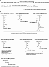
Mono-ADP-ribosylation cycle and the corresponding products of protein mono-ADP-ribosylation. (A) Mono-ADP-ribosyl transferases catalyze the transfer of the ADP-ribose moiety of NAD+ to an acceptor molecule (free amino acids, proteins, DNA, and RNA, etc.). The action of mono-ADP-ribosyl-amino acid hydrolases, which regenerate the corresponding free acceptor molecule, is consistent with the presence of a mono-ADP-ribosylation cycle. (B) The transferase-catalyzed reaction of protein mono-ADP-ribosylation results in a stereo-specific formation of Ω-N-(C-1-ADP-ribosyl)-l-arginine, ɛ-N-(C-1-ADP-ribosyl)-l-asparagine, ω-N-(C-1-ADP-ribosyl)-l-diphtamide, γ-S-(C-1-ADP-ribosyl)-l-cysteine, ɛ-O-(C-1-ADP-ribosyl)-l-glutamate, δ-O-(C-1-ADP-ribosyl)-l-aspartate, or O-(ADP-ribosyl)-l-phosphoserine.
FIG. 3.
FIG. 3.
Domain structures of the human Pl-MART family. A new classification and a schematic comparison of protein structures of the 11 members of the Pl-MART family, based on the literature and database searches, are shown. The most significant domains detected have been indicated. The WWE domain is named after three of its conserved residues (W/W/E) and is predicted to mediate specific protein-protein interactions in ubiquitin- and ADP-ribose conjugation systems (24). Although the exact roles of the conserved macroH2A/A1pp domains remain unknown, they have been proposed to have ADP-ribose 1"-phosphate (Appr-1"p)-processing activity and may regulate mono-ADP-ribosylation (219). ZF, C3H-type zinc finger domain; RRM, RNA recognition motif (252); UIM, ubiquitin interaction motif (22); MVP-ID, M-vault particle interaction domain; TPH, Ti-PARP homologous domain; GRD, glycine-rich domain.
FIG. 4.
FIG. 4.
Possible metabolism of acetyl-ADP-ribose (O-AADPR) and mono-ADP-ribosylation of proteins by SIRTs. SIRTs cleave the glycosidic bond between the Nam and ADP-ribose portions of NAD+. The ADP-ribose intermediate is necessary for the deacetylation reaction. Following hydrolysis of the glycosidic bond, nicotinamide is released, and ADP-ribose binds the acetyl-peptide, forming of an O-alkylamidate intermediate. The acetyl group removed from the target substrate is transferred to the ADP-ribose moiety to form 2′-acetyl-ADP-ribose (2′-O-AADPR) and then subsequently released together with the deacetylated protein from the enzyme-intermediate complex. 2′-O-AADPR spontaneously equilibrates with the regioisomer 3′-acetyl-ADP-ribose (3′-O-AADPR) through trans-esterification. In mammalian cells, acetyl-ADP-ribose can be deacetylated by esterases to the ATP precursor ADP-ribose or can function as an acetyl donor to acetylate unknown substrates by nuclear trans-acetylases. Transformation of acetyl-ADP-ribose into AMP and acetyl-ribose-5-phosphate by ADP-ribose hydrolases of the Nudix family is not clearly established. The possible deacetylation-dependent and -independent transfers of mono-ADP-ribose to proteins catalyzed by SIRTs are shown in the lower part of the figure.
FIG. 5.
FIG. 5.
Poly-ADP-ribose metabolism. Steps 1 to 3 and steps 4 to 7 of the poly-ADP-ribose cycle represent the anabolic and catabolic reactions, respectively, in the metabolism of poly-ADP-ribose. The synthesis of poly-ADP-ribose requires three distinct PARP activities: step 1, initiation or mono-ADP-ribosylation of a specific glutamic (?) acid residue(s) in the corresponding PARP enzyme (acceptor); step 2, elongation of the polymer; and step 3, branching of the polymer. The degradation requires at least four (alternative) PARG and (P/M)ARH activities: step 4, exoglycosidase and endoglycosidase (PARG) activities, respectively, that hydrolyze the glycosidic linkages between the ADP-ribose units; step 5, potential poly-ADP-ribosyl-protein hydrolase activities; and step 6, MARH, or step 7, mono-ADP-ribosyl-protein lyase activities. Chemical structures in this figure were drawn with MarvinSketch, version 4.0.4 (ChemAxon, Budapest, Hungary).
FIG. 6.
FIG. 6.
Domain structures of the human PARP family. A classification and a schematic comparison of protein structures of the six members of the bona fide PARP family, based on literature and database searches, are shown. The most significant domains detected have been indicated. The PRD domain is called the PARP regulatory domain and may be involved in regulation of the PARP-branching activity. The WGR domain is named after the most conserved central motif (W/G/R) of the domain. This motif is found in a variety of poly(A) polymerases and other proteins of unknown function. The BRCT domain is named after the breast cancer suppressor protein-1 (BRCA1) carboxy-terminal domain and is found within many DNA damage repair and cell cycle checkpoint proteins (446). The unique diversity of this domain superfamily allows BRCT modules to interact by forming homo- or hetero-BRCT multimers and phosphorylation-dependent BRCT-non-BRCT interactions (139, 446). The sterile alpha motif (SAM) is a widespread domain in signaling and nuclear proteins and mediates homo- or heterodimerization in many cases (reviewed in reference 20). The ankyrin repeat domains (ARD) mediate protein-protein interactions in very diverse families of proteins (279). The number of ankyrin repeats in a protein can range from 2 to over 20 (279). The vault protein inter-alpha-trypsin (VIT) and von Willebrand type A (vWA) domains are conserved domains found in all inter-alpha-trypsin inhibitor (ITI) family members (261). Although the exact roles of these domains remain unknown, they are presumed to be involved in mediating protein-protein interactions (261). ZF-I and ZF-II, PARP-1-type zinc finger domains (they can act as DNA nick sensors and general DNA-binding domains [161]); SAP, SAF/Acinus/PIAS-DNA-binding domain; LZM, putative leucine zipper-like motif; MVP-ID, major-vault particle interaction domain; NLS, nuclear localization signal; CLS, centriole-localization signal; HPS, His-Pro-Ser region.
FIG. 7.
FIG. 7.
Domain structures of the human PARG isoforms. A classification and a schematic comparison of protein structures of the four PARG isoforms is shown (data are from references , , and 315). NLS, nuclear localization signals (amino acids 10 to 16, 32 to 38, 421 to 446, and 838 to 844); NES, nuclear export signals (amino acids 126 to 134, 421 to 446, and 881 to 888). Active sites: E728, E738, E756, E757, and T995.
FIG. 8.
FIG. 8.
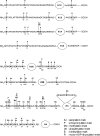
Summary of known histone modifications in the human linker and core histones. The covalent modifications on histones include acetylation, phosphorylation, methylation, ubiquitination, biotinylation, and mono-ADP-ribosylation (data are from references , , , , , , , , , , and 430).

Similar articles

Cited by

References

    1. Adamietz, P., and A. Rudolph. 1984. ADP-ribosylation of nuclear proteins in vivo. Identification of histone H2B as a major acceptor for mono- and poly(ADP-ribose) in dimethyl sulfate-treated hepatoma AH 7974 cells. J. Biol. Chem. 259:6841-6846. - PubMed
    1. Adebanjo, O. A., H. K. Anandatheerthavarada, A. P. Koval, B. S. Moonga, G. Biswas, L. Sun, B. R. Sodam, P. J. Bevis, C. L. Huang, S. Epstein, F. A. Lai, N. G. Avadhani, and M. Zaidi. 1999. A new function for CD38/ADP-ribosyl cyclase in nuclear Ca2+ homeostasis. Nat. Cell Biol. 1:409-414. - PubMed
    1. Adolph, K. W., and M. K. Song. 1985. Variations in ADP-ribosylation of nuclear scaffold proteins during the HeLa cell cycle. Biochem. Biophys. Res. Commun. 126:840-847. - PubMed
    1. Adriouch, S., W. Ohlrogge, F. Haag, F. Koch-Nolte, and M. Seman. 2001. Rapid induction of naive T cell apoptosis by ecto-nicotinamide adenine dinucleotide: requirement for mono(ADP-ribosyl)transferase 2 and a downstream effector. J. Immunol. 167:196-203. - PubMed
    1. Aguiar, R. C., K. Takeyama, C. He, K. Kreinbrink, and M. A. Shipp. 2005. B-aggressive lymphoma family proteins have unique domains that modulate transcription and exhibit poly(ADP-ribose) polymerase activity. J. Biol. Chem. 280:33756-33765. - PubMed

Publication types

MeSH terms

LinkOut - more resources