. 2006 Sep;13(9):981-90.
doi: 10.1128/CVI.00156-06.

Distinct contributions of vaccine-induced immunoglobulin G1 (IgG1) and IgG2a antibodies to protective immunity against influenza

Affiliations

Distinct contributions of vaccine-induced immunoglobulin G1 (IgG1) and IgG2a antibodies to protective immunity against influenza

Victor C Huber et al. Clin Vaccine Immunol. 2006 Sep.

Abstract

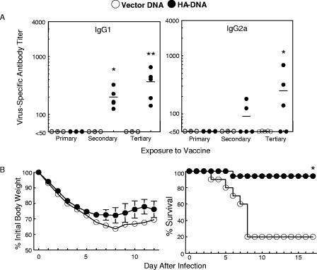
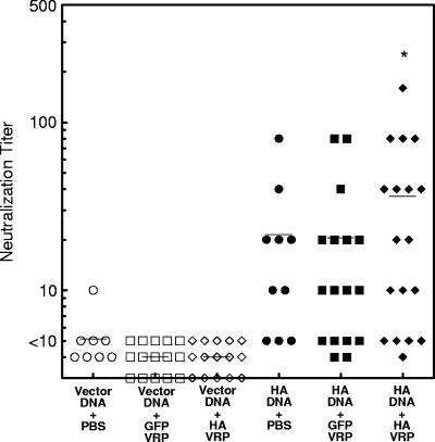
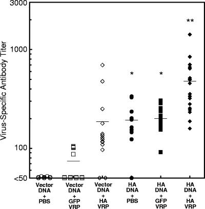
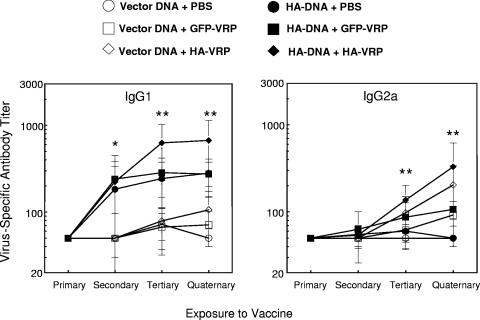
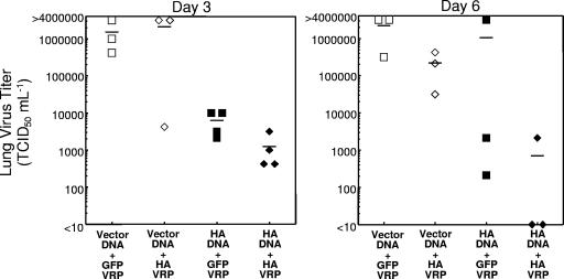
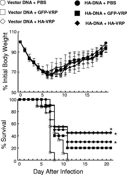
Vaccination represents the most effective form of protection against influenza infection. While neutralizing antibodies are typically measured as a correlate of vaccine-induced protective immunity against influenza, nonneutralizing antibodies may contribute to protection or amelioration of disease. The goal of this study was to dissect the individual contributions of the immunoglobulin G1 (IgG1) and IgG2a antibody isotypes to vaccine-induced immunity against influenza virus. To accomplish this, we utilized an influenza vaccine regimen that selectively enhanced IgG1 or IgG2a antibodies by using either DNA or viral replicon particle (VRP) vectors expressing influenza virus hemagglutinin (HA) (HA-DNA or HA-VRP, respectively). After HA-DNA vaccination, neutralizing antibodies were detected by both in vitro (microneutralization) and in vivo (lung viral titer) methods and were associated with increased IgG1 expression by enzyme-linked immunosorbent assay (ELISA). Vaccination with HA-VRP did not strongly stimulate either neutralizing or IgG1 antibodies but did induce IgG2a antibodies. Expression of IgG2a antibodies in this context correlated with clearance of virus and increased protection against lethal influenza challenge. Increased induction of both antibody isotypes as measured by ELISA was a better correlate for vaccine efficacy than neutralization alone. This study details separate but important roles for both IgG1 and IgG2a expression in vaccination against influenza and argues for the development of vaccine regimens that stimulate and measure expression of both antibody isotypes.


Figures

FIG. 1.
FIG. 1.
Response of BALB/c mice to HA-DNA gene gun vaccination and influenza challenge. (A) IgG1 and IgG2a antibody levels were measured by ELISA 14 days after primary, secondary, and tertiary exposures to vector control or influenza HA delivered by DNA vaccine at 3-week intervals. Data are reported for six vector control-inoculated mice, with the exception of IgG2a readings at day 14 of the tertiary response (five mice). Data are shown for six HA-inoculated mice for the primary response and five HA-inoculated mice for the secondary and tertiary responses for both isotypes. An asterisk indicates a significant difference in titer compared to that for mice inoculated with vector DNA (P < 0.01 by ANOVA). A double asterisk indicates a significant difference in titer compared to those for all other groups (P < 0.01 by ANOVA). (B) Mice were challenged with 3 MLD50 HK/Syd on day 21 of the tertiary response to the vaccine. Mean levels of weight loss ± standard deviations are pictured for seven randomly selected mice per group. Survival data are reported for 10 vector control mice and 17 HA-vaccinated mice. An asterisk indicates a significant difference in survival compared to that of controls (P < 0.01 by log rank test of the Kaplan-Meier survival data).
FIG. 2.
FIG. 2.
Serum neutralizing antibody response of BALB/c mice to DNA prime followed by VRP boost. Microneutralization titers against HK/Syd (2,000 TCID50 ml−1) at day 21 of the quaternary response to the vaccine are reported. Data are reported for the following numbers of mice in the various groups: for vector DNA plus PBS, n = 8; for vector DNA plus GFP-VRP, n = 15; for vector DNA plus HA-VRP, n = 15; for HA-DNA plus PBS, n = 10; for HA-DNA plus GFP-VRP, n = 17; and for HA-DNA plus HA-VRP, n = 18. An asterisk indicates a significant difference in titer compared to those for groups inoculated with vector DNA (P < 0.01 by ANOVA).
FIG. 3.
FIG. 3.
Virus-specific serum antibody response of BALB/c mice to DNA prime followed by VRP boost. IgG antibody titers against HK/Syd (1 μg HA ml−1) were measured by ELISA and are shown at day 21 of the quaternary response to the vaccine. Data are reported for the following numbers of mice in the various groups: for vector DNA plus PBS, n = 8; for vector DNA plus GFP-VRP, n = 15; for vector DNA plus HA-VRP, n = 15; for HA-DNA plus PBS, n = 10; for HA-DNA plus GFP-VRP, n = 17; and for HA-DNA plus HA-VRP, n = 18. An asterisk indicates a significant difference in titer for mice vaccinated with HA-DNA plus PBS and mice vaccinated with HA-DNA plus GFP-VRP compared to those for mice in groups inoculated with vector DNA plus PBS and vector DNA plus GFP-VRP (P < 0.01 by ANOVA). A double asterisk indicates a significant difference in titer for mice vaccinated with HA-DNA plus HA-VRP compared to those for all three groups of vector DNA-vaccinated mice (P < 0.01 by ANOVA).
FIG. 4.
FIG. 4.
Virus-specific serum antibody response of BALB/c mice to DNA prime followed by VRP boost. IgG1 and IgG2a antibody isotype titers against HK/Syd (1 μg HA ml−1) were measured by ELISA and are shown after primary (day 14), secondary (day 14), tertiary (day 21), and quaternary (day 21) responses to the vaccine. For all days and isotypes measured, the following numbers of mice were included in the various groups: for vector DNA plus PBS, n = 8; for vector DNA plus GFP-VRP, n = 15; for vector DNA plus HA-VRP, n = 15; for HA-DNA plus PBS, n = 10; for HA-DNA plus GFP-VRP, n = 17; and for HA-DNA plus HA-VRP, n = 18. An asterisk indicates a significant difference in titer for HA-DNA-vaccinated mice compared to that for vector DNA-vaccinated mice (P < 0.01 by ANOVA). A double asterisk indicates a significant difference in titer for mice vaccinated with HA-DNA plus HA-VRP compared to those for all other groups (P < 0.01 by ANOVA).
FIG. 5.
FIG. 5.
Lung viral titers of BALB/c mice after HK/Syd challenge. After the quaternary exposure to the vaccine, mice were infected with 100 MID50 HK/Syd, and lung viral titers at days 3 and 6 after inoculation were determined. Data are reported for the following numbers of mice in the various groups: for vector DNA plus GFP-VRP, n = 3; for vector DNA plus HA-VRP, n = 3; for HA-DNA plus GFP-VRP, n = 4; and for HA-DNA plus HA-VRP, n = 4. On day 6 after viral challenge, all four groups contained three mice each.
FIG. 6.
FIG. 6.
Survival of BALB/c mice after HK/Syd challenge. After the quaternary exposure to the vaccine, mice were infected with 10 MLD50 HK/Syd and monitored for morbidity (percent weight loss) (top) and mortality (percent survival) (bottom). Data are reported for the following numbers of mice in the various groups: for vector DNA plus PBS, n = 8; for vector DNA plus GFP-VRP, n = 9; for vector DNA plus HA-VRP, n = 9; for HA-DNA plus PBS, n = 10; for HA-DNA plus GFP-VRP, n = 10; and for HA-DNA plus HA-VRP, n = 11. An asterisk indicates a significant difference in results compared to those for vector DNA-plus-PBS and vector DNA-plus-GFP-VRP groups (P < 0.05 by log rank test of the Kaplan-Meier survival data).
FIG. 7.
FIG. 7.
Effector CD8+ T-cell populations in the BAL fluid and livers of BALB/c mice after HK/Syd challenge. After the quaternary response to the vaccine, mice were infected with 100 MID50 HK/Syd. BAL fluid and liver cells were isolated and analyzed for CD8+ T-cell populations on day 6 after challenge. All groups consisted of three mice.

References

    1. Arulanandam, B. P., M. O'Toole, and D. W. Metzger. 1999. Intranasal interleukin-12 is a powerful adjuvant for protective mucosal immunity. J. Infect. Dis. 180:940-949. - PubMed
    1. Barackman, J. D., G. Ott, and D. T. O'Hagan. 1999. Intranasal immunization of mice with influenza vaccine in combination with the adjuvant LT-R72 induces potent mucosal and serum immunity which is stronger than that with traditional intramuscular immunization. Infect. Immun. 67:4276-4279. - PMC - PubMed
    1. Beigel, J. H., J. Farrar, A. M. Han, F. G. Hayden, R. Hyer, M. D. de Jong, S. Lochindarat, T. K. Nguyen, T. H. Nguyen, T. H. Tran, A. Nicoll, S. Touch, K. Y. Yuen, et al. 2005. Avian influenza A (H5N1) infection in humans. N. Engl. J. Med. 353:1374-1385. - PubMed
    1. Belshe, R. B., W. C. Gruber, P. M. Mendelman, H. B. Mehta, K. Mahmood, K. Reisinger, J. Treanor, K. Zangwill, F. G. Hayden, D. I. Bernstein, K. Kotloff, J. King, P. A. Piedra, S. L. Block, L. Yan, and M. Wolff. 2000. Correlates of immune protection induced by live, attenuated, cold-adapted, trivalent, intranasal influenza virus vaccine. J. Infect. Dis. 181:1133-1137. - PubMed
    1. Benne, C. A., M. Harmsen, W. van der Graaff, A. F. Verheul, H. Snippe, and C. A. Kraaijeveld. 1997. Influenza virus neutralizing antibodies and IgG isotype profiles after immunization of mice with influenza A subunit vaccine using various adjuvants. Vaccine 15:1039-1044. - PubMed

Publication types

MeSH terms