

Release of plasmid DNA from intravascular stents coated with ultrathin multilayered polyelectrolyte films
- PMID: 16961308
- PMCID: PMC2522324
- DOI: 10.1021/bm0604808
Release of plasmid DNA from intravascular stents coated with ultrathin multilayered polyelectrolyte films
Abstract
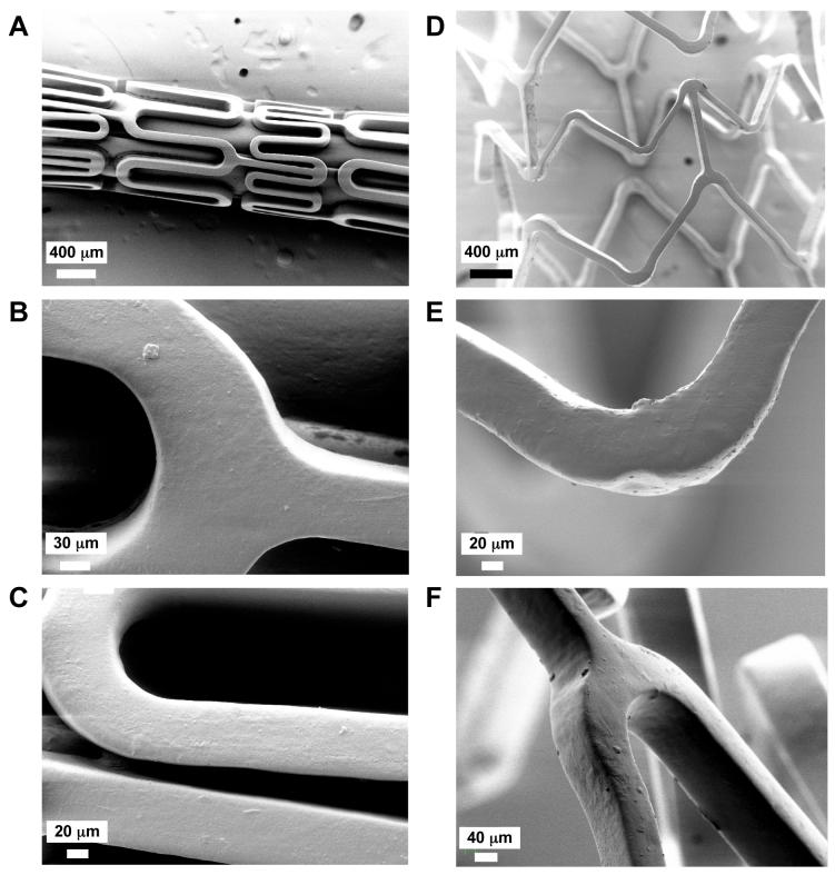
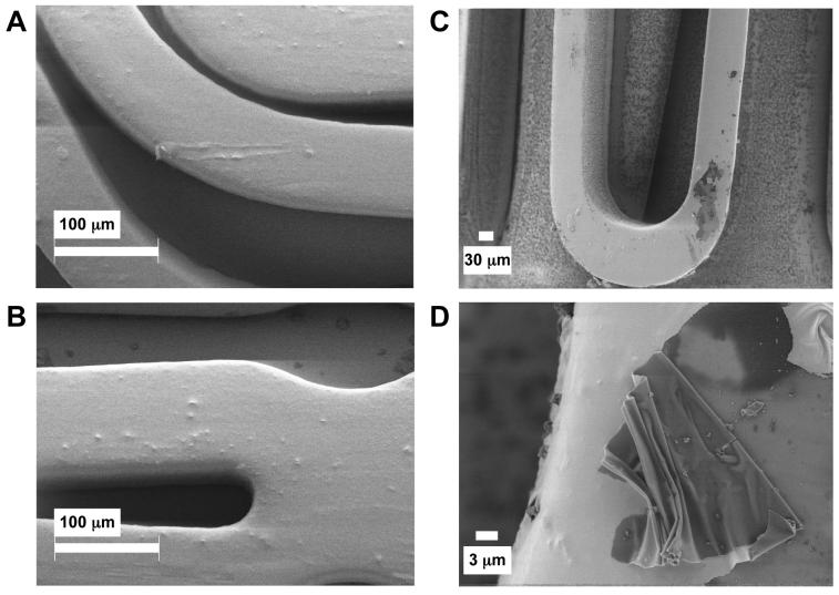
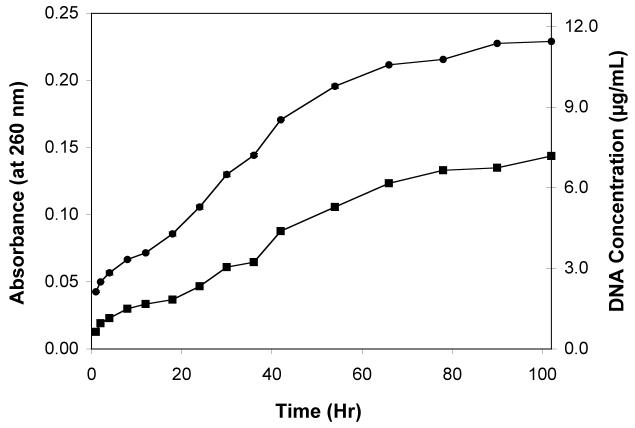
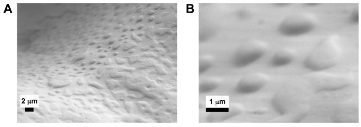
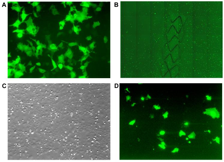
Materials that permit control over the release of DNA from the surfaces of topologically complex implantable devices, such as intravascular stents, could contribute to the development of new approaches to the localized delivery of DNA. We report the fabrication of ultrathin, multilayered polyelectrolyte films that permit both the immobilization and controlled release of plasmid DNA from the surfaces of stainless steel intravascular stents. Our approach makes use of an aqueous-based, layer-by-layer method for the assembly of nanostructured thin films consisting of alternating layers of plasmid DNA and a hydrolytically degradable polyamine. Characterization of coated stents using scanning electron microscopy (SEM) demonstrated that stents were coated uniformly with an ultrathin film ca. 120 nm thick that adhered conformally to the surfaces of stent struts. These ultrathin films did not crack, peel, or delaminate substantially from the surface after exposure to a range of mechanical challenges representative of those encountered during stent deployment (e.g., balloon expansion). Stents coated with eight bilayers of degradable polyamine and a plasmid encoding enhanced green fluorescent protein (EGFP) sustained the release of DNA into solution for up to four days when incubated in phosphate buffered saline at 37 degrees C, and coated stents were capable of mediating the expression of EGFP in a mammalian cell line without the aid of additional transfection agents. The approach reported here could, with further development, contribute to the development of localized gene-based approaches to the treatment of cardiovascular diseases or related conditions.
Figures
Similar articles
-
Multilayered polyelectrolyte assemblies as platforms for the delivery of DNA and other nucleic acid-based therapeutics.Adv Drug Deliv Rev. 2008 Jun 10;60(9):979-99. doi: 10.1016/j.addr.2008.02.010. Epub 2008 Mar 4. Adv Drug Deliv Rev. 2008. PMID: 18395291 Free PMC article. Review.
-
Assembly of erodible, DNA-containing thin films on the surfaces of polymer microparticles: toward a layer-by-layer approach to the delivery of DNA to antigen-presenting cells.Acta Biomater. 2009 Mar;5(3):913-24. doi: 10.1016/j.actbio.2008.08.022. Epub 2008 Sep 11. Acta Biomater. 2009. PMID: 18838346 Free PMC article.
-
Multilayered films fabricated from plasmid DNA and a side-chain functionalized poly(beta-amino ester): surface-type erosion and sequential release of multiple plasmid constructs from surfaces.Langmuir. 2007 Oct 23;23(22):11139-46. doi: 10.1021/la702021s. Epub 2007 Sep 22. Langmuir. 2007. PMID: 17887783
-
Polyelectrolyte multilayers promote stent-mediated delivery of DNA to vascular tissue.Biomacromolecules. 2013 May 13;14(5):1696-704. doi: 10.1021/bm4005222. Epub 2013 May 2. Biomacromolecules. 2013. PMID: 23597075 Free PMC article.
-
Delivery of plasmid DNA to vascular tissue in vivo using catheter balloons coated with polyelectrolyte multilayers.Biomaterials. 2011 Jan;32(2):610-8. doi: 10.1016/j.biomaterials.2010.09.009. Epub 2010 Oct 8. Biomaterials. 2011. PMID: 20933275 Free PMC article.
Cited by
-
Polymer multilayer tattooing for enhanced DNA vaccination.Nat Mater. 2013 Apr;12(4):367-76. doi: 10.1038/nmat3550. Epub 2013 Jan 27. Nat Mater. 2013. PMID: 23353628 Free PMC article.
-
Controlling the surface-mediated release of DNA using 'mixed multilayers'.Bioeng Transl Med. 2016 Jun;1(2):181-192. doi: 10.1002/btm2.10023. Epub 2016 Aug 26. Bioeng Transl Med. 2016. PMID: 27981243 Free PMC article.
-
Multilayered polyelectrolyte assemblies as platforms for the delivery of DNA and other nucleic acid-based therapeutics.Adv Drug Deliv Rev. 2008 Jun 10;60(9):979-99. doi: 10.1016/j.addr.2008.02.010. Epub 2008 Mar 4. Adv Drug Deliv Rev. 2008. PMID: 18395291 Free PMC article. Review.
-
Cross-linked bioreducible layer-by-layer films for increased cell adhesion and transgene expression.J Phys Chem B. 2010 Apr 29;114(16):5283-91. doi: 10.1021/jp100486h. J Phys Chem B. 2010. PMID: 20369813 Free PMC article.
-
Polymer Multilayers that Promote the Rapid Release and Contact Transfer of DNA.Biomacromolecules. 2015 Sep 14;16(9):2998-3007. doi: 10.1021/acs.biomac.5b00905. Epub 2015 Aug 28. Biomacromolecules. 2015. PMID: 26285737 Free PMC article.
References
-
- Morice MC, Serruys PW, Sousa JE, Fajadet J, Ban Hayashi E, Perin M, Colombo A, Schuler G, Barragan P, Guagliumi G, Molnar F, Falotico R. N Engl J Med. 2002;346:1773–80. - PubMed
-
- Moses JW, Leon MB, Popma JJ, Fitzgerald PJ, Holmes DR, O’Shaughnessy C, Caputo RP, Kereiakes DJ, Williams DO, Teirstein PS, Jaeger JL, Kuntz RE. N Engl J Med. 2003;349:1315–23. - PubMed
-
- Sousa JE, Serruys PW, Costa MA. Circulation. 2003;107:2383–9. - PubMed
-
- Tanabe K, Regar E, Lee CH, Hoye A, van der Giessen WJ, Serruys PW. Curr Pharm Des. 2004;10:357–67. - PubMed
-
- Stone GW, Ellis SG, Cox DA, Hermiller J, O’Shaughnessy C, Mann JT, Turco M, Caputo R, Bergin P, Greenberg J, Popma JJ, Russell ME. N Engl J Med. 2004;350:221–31. - PubMed
Publication types
MeSH terms
Substances
Grants and funding
LinkOut - more resources
Full Text Sources
Other Literature Sources
