. 2006 Sep 8:3:13.
doi: 10.1186/1743-8977-3-13.

Translocation and potential neurological effects of fine and ultrafine particles a critical update

Affiliations

Translocation and potential neurological effects of fine and ultrafine particles a critical update

Annette Peters et al. Part Fibre Toxicol. .

Abstract

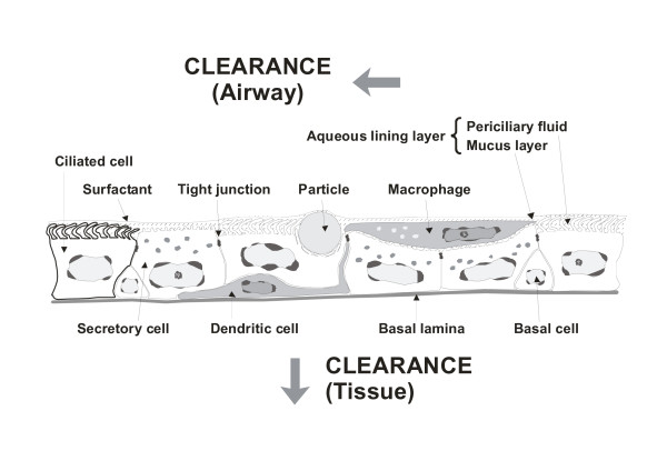
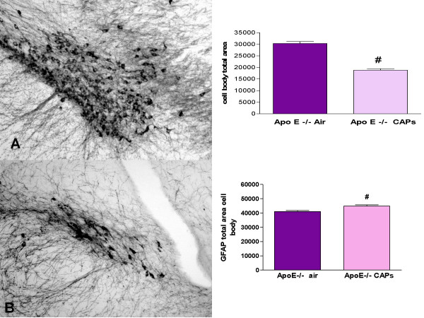
Particulate air pollution has been associated with respiratory and cardiovascular disease. Evidence for cardiovascular and neurodegenerative effects of ambient particles was reviewed as part of a workshop. The purpose of this critical update is to summarize the evidence presented for the mechanisms involved in the translocation of particles from the lung to other organs and to highlight the potential of particles to cause neurodegenerative effects. Fine and ultrafine particles, after deposition on the surfactant film at the air-liquid interface, are displaced by surface forces exerted on them by surfactant film and may then interact with primary target cells upon this displacement. Ultrafine and fine particles can then penetrate through the different tissue compartments of the lungs and eventually reach the capillaries and circulating cells or constituents, e.g. erythrocytes. These particles are then translocated by the circulation to other organs including the liver, the spleen, the kidneys, the heart and the brain, where they may be deposited. It remains to be shown by which mechanisms ultrafine particles penetrate through pulmonary tissue and enter capillaries. In addition to translocation of ultrafine particles through the tissue, fine and coarse particles may be phagocytized by macrophages and dendritic cells which may carry the particles to lymph nodes in the lung or to those closely associated with the lungs. There is the potential for neurodegenerative consequence of particle entry to the brain. Histological evidence of neurodegeneration has been reported in both canine and human brains exposed to high ambient PM levels, suggesting the potential for neurotoxic consequences of PM-CNS entry. PM mediated damage may be caused by the oxidative stress pathway. Thus, oxidative stress due to nutrition, age, genetics among others may increase the susceptibility for neurodegenerative diseases. The relationship between PM exposure and CNS degeneration can also be detected under controlled experimental conditions. Transgenic mice (Apo E -/-), known to have high base line levels of oxidative stress, were exposed by inhalation to well characterized, concentrated ambient air pollution. Morphometric analysis of the CNS indicated unequivocally that the brain is a critical target for PM exposure and implicated oxidative stress as a predisposing factor that links PM exposure and susceptibility to neurodegeneration. Together, these data present evidence for potential translocation of ambient particles on organs distant from the lung and the neurodegenerative consequences of exposure to air pollutants.


Figures

Figure 1
Figure 1
Schematic drawing of airway epithelial barrier with macrophages and dendritic cells, exposed to a fine particle (redrawn after McWilliam et al., 2000).
Figure 2
Figure 2
COX2 expression in frontal cortex and hippocampus. (A and D) COX2 mRNA abundance was measured by RT-PCR and normalized for 18s rRNA levels. Means ± SEMs are shown. COX2 mRNA was significantly elevated in the high exposure group in both frontal cortex (A, * p = 0.009) and hippocampus (D, * p = 0.04) from the high exposure group. (B and E) COX2 protein expression in sections of paraffin-embedded tissues was localized by COX2 immunohistochemistry (IHC) and the percent of tissue area that was immunoreactive (COX2 IR) was measured by quantitative image analysis. Means ± SEMs are shown. COX2 IR was significantly elevated in frontal cortex (B, * p = 0.01), but not in hippocampus (E) from the high exposure group. (C) Representative COX IHC in frontal cortex from a subject in the high exposure group showing strong staining of endothelial cells in the capillaries (*), and pyramidal neurons (arrow), while other neurons were negative (arrowheads). Scale = 20 μm. (F) Representative COX IHC in dentate gyrus from a subject in the high exposure group showing COX2 positive neurons (arrowheads) and capillaries (short arrow). Scale = 15 μm.
Figure 3
Figure 3
Aβ42 accumulation in frontal cortex and hippocampus. Aβ42 was localized in sections of paraffin-embedded tissues by IHC. (A) Aβ42 IHC stained pyramidal neurons (p), astrocytes (arrows) and astrocytic processes (arrowheads) around blood vessels (*). (B) In addition to accumulation in pyramidal neurons (p) Aβ42 was deposited in smooth muscle cells (arrows) in cortical arterioles (*). A dead neuron surrounded by glial cells is indicated (arrowhead). (C and D) Quantitative image analysis of Aβ42 IHC showed a significant increase in Aβ42 immunoreactivity (Aβ42 IR) in both frontal cortex (C, * p = 0.04) and hippocampus (D, * p = 0.001) in the high exposure group. (E) Aβ42 IHC of frontal cortex from a 38 year old subject from Mexico City showing diffuse plaque-like staining with surrounding reactive astrocytes (arrows). Scale = 20 μm.
Figure 4
Figure 4
Micrographs of the SN of (A) Air and (B) CAPs exposed Apo E -/- mouse brain and representative examples of the nucleus compacta which house the dopaminergic neurons, vulnerable to OS. Sections were taken from the same brain level. Neurons and astrocytes were immunocytochemical stained with TH and GFAP, respectively. A 29% reduction of TH staining and an 8% increase in GFAP was measured in the Apo E-/- PM exposed mice relative to air exposed Apo E-/- No differences in TH or GFAP bodies were observed in C57 Air or PM exposed animals. Sections 300× magnification.
Figure 5
Figure 5
Genomics: BV2 microglia were exposed in triplicate (2 hr) to CAPs (75 μg/ml), and other chemicals known to produce OS (i.e., LPS (2.5 ng/ml); H202 (0.2 mM), Diesel exhaust filtrate (100 μg/ml). RNAs from each test group were analyzed on Affymetrix universal microarrays. Relative to the other treatment and controls, CAPs produced significant increases/decreases in genes coding for apoptotic, TNF and Toll receptors; cytokines, interferons, kinase transcription, oncogenes and growth factors.

References

    1. Brunekreef B, Holgate ST. Air pollution and health. Lancet. 2002;360:1233–1242. doi: 10.1016/S0140-6736(02)11274-8. - DOI - PubMed
    1. Brook RD, Franklin B, Cascio WE, Hong Y, Howard G, Lipsett M, Luepker RV, Mittleman MA, Samet JM, Smith SCJ, Tager IB. Air Pollution and Cardiovascular Disease: A statement of the health care professionals from the expert panel on population and prevention science of the American Heart Association. Circulation. 2004;109:2655–2671. doi: 10.1161/01.CIR.0000128587.30041.C8. - DOI - PubMed
    1. Kappos AD, Bruckmann P, Eikmann T, Englert N, Heinrich U, Hoppe P, Koch E, Krause GH, Kreyling WG, Rauchfuss K, Rombout P, Schulz-Klemp V, Thiel WR, Wichmann HE. Health effects of particles in ambient air. Int J Hyg Environ Health. 2004;207:399–407. doi: 10.1078/1438-4639-00306. - DOI - PubMed
    1. Ibald-Mulli A, Wichmann H, Kreyling WG, Peters A. Epidemiological Evidence on Health Effects of Ultrafine Particles. Journal of Aerosol Medicine. 2002;15:189–201. doi: 10.1089/089426802320282310. - DOI - PubMed
    1. Schulz H, Harder V, Ibald-Mulli A, Khandoga A, Koenig W, Krombach F, Radykewicz R, Stampfl A, Thorand B, Peters A. Cardiovascular effects of fine and ultrafine particles. J Aerosol Med. 2005;18:1–22. doi: 10.1089/jam.2005.18.1. - DOI - PubMed