Case Reports
. 2006 Sep 10:6:33.
doi: 10.1186/1471-2377-6-33.

Extrapontine myelinolysis presenting as acute parkinsonism

Affiliations
Case Reports

Extrapontine myelinolysis presenting as acute parkinsonism

J Sajith et al. BMC Neurol. .

Abstract

Background: Extrapontine myelinolysis presenting with extra pyramidal features suggestive of parkinsonism may be a challenging clinical syndrome. Clinicians should maintain their vigilance while correcting electrolyte imbalances, especially with associated co-morbidity.

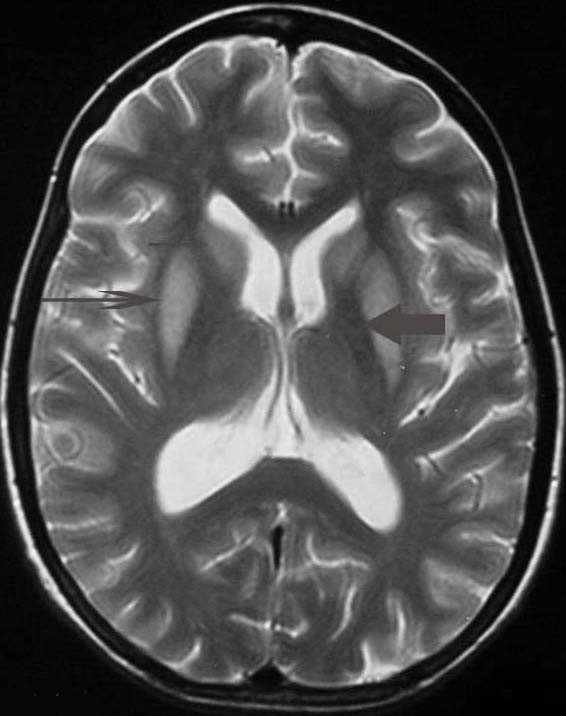
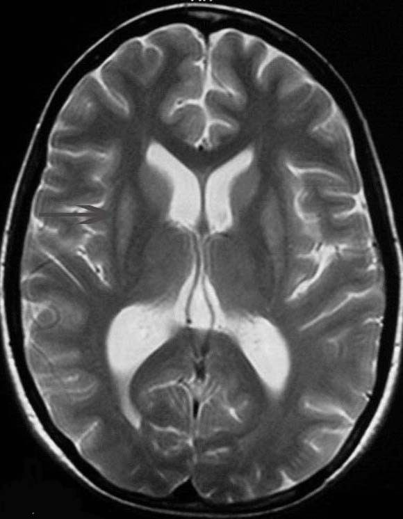
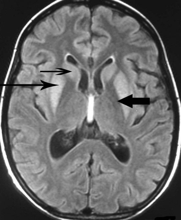
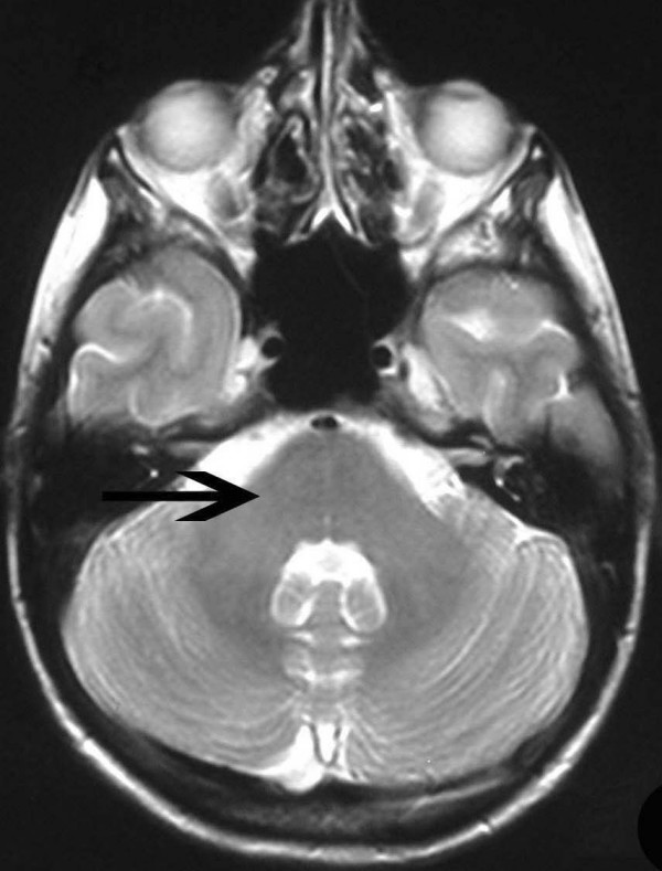
Case presentation: A 41-year-old woman presented with acute parkinsonism like features while on a holiday. This followed slow correction of hyponatraemia after repeated vomiting. MRI changes were suggestive of Extrapontine myelinolysis(EPM). This case is at variance with four previous cases reported in the medical literature in that the patient made a full clinical recovery and the MR changes resolved with symptomatic support alone.

Conclusion: Extrapontine myelinolysis could make a complete recovery with symptomatic support alone. During hyponatraemia correction, rapid osmotic shifts of fluid that cause hypernatremia, causes myelinolysis rather than absolute serum sodium level. Even gradual correction of hyponatraemia can produce myelinolysis, especially with pre-existing malnourishment, alcoholism, drug misuse, Addison's disease and immuno-suppression. Pallidial sparing is typical of EPM in MRI scans.


Figures

Figure 1
Figure 1
T2 weighted magnetic resonance scan image showing bilaterally symmetrical hyperintensities in Caudate nucleus (small, thin arrow), Putamen (long arrow), with sparing of Globus Pallidus (broad arrow), suggestive of Extrapontine myelinolysis.
Figure 2
Figure 2
T2 weighted magnetic resonance scan image after two months showing marked improvement, mainly in putamen (long arrow).
Figure 3
Figure 3
T2 weighted FLAIR images of magnetic resonance scan showing bilaterally symmetrical hyperintensities in Caudate nucleus (small, thin arrow), Putamen (long arrow), with sparing of Globus Pallidus (broad arrow), suggestive of Extrapontine myelinolysis.
Figure 4
Figure 4
T2 weighted magnetic resonance scan image of pons showing absolutely no changes.

Similar articles

Cited by

References

    1. Tomita I, Satoh H, Satoh A, Seto M, Tsujihata M, Yoshimura T. Extrapontine myelinolysis presenting with parkinsonism as a sequel of rapid correction of hyponatraemia. JNNP. 1997;62:422–423. - PMC - PubMed
    1. Seiser A, Schwarz S, Aichinger-Steiner M, Funk G, Schnider P, Brainin M. Parkinsonism and dystonia in central pontine and extrapontine myelinolysis. JNNP. 1998;65:119–121. - PMC - PubMed
    1. Chu K, Kang D-W, Ko S-B, Kim M. Diffusion-weighted MR findings of central pontine and extrapontine myelinolysis. ActaNeurol Scand. 2001;104:385–388. doi: 10.1034/j.1600-0404.2001.00096.x. - DOI - PubMed
    1. Schimrigk S, Amoiridis G. Extrapontine myelinolysis. JNNP. 1996;61:250–251. - PMC - PubMed
    1. Martin RJ. Central pontine and extrapontine myelinolysis:the osmotic demyelination syndromes. J Neurol Neurosurg Psychiatry. 2004;75:iii22–8. - PMC - PubMed

Publication types

LinkOut - more resources