

Emergence of physiological rhythmicity in term and preterm neonates in a neonatal intensive care unit
- PMID: 16961937
- PMCID: PMC1574348
- DOI: 10.1186/1740-3391-4-11
Emergence of physiological rhythmicity in term and preterm neonates in a neonatal intensive care unit
Abstract
Background: Biological rhythmicity, particularly circadian rhythmicity, is considered to be a key mechanism in the maintenance of physiological function. Very little is known, however, about biological rhythmicity pattern in preterm and term neonates in neonatal intensive care units (NICU). In this study, we investigated whether term and preterm neonates admitted to NICU exhibit biological rhythmicity during the neonatal period.
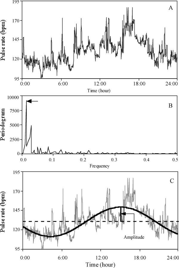
Methods: Twenty-four-hour continuous recording of four physiological variables (heart rate: HR recorded by electrocardiogram; pulse rate: PR recorded by pulse oxymetry; respiratory rate: RR; and oxygen saturation of pulse oxymetry: SpO2) was conducted on 187 neonates in NICU during 0-21 days of postnatal age (PNA). Rhythmicity was analyzed by spectral analysis (SPSS procedure Spectra). The Fisher test was performed to test the statistical significance of the cycles. The cycle with the largest peak of the periodogram intensities was determined as dominant cycle and confirmed by Fourier analysis. The amplitudes and amplitude indexes for each dominant cycle were calculated.
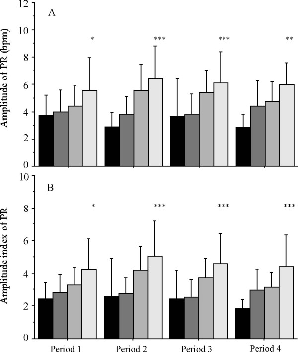
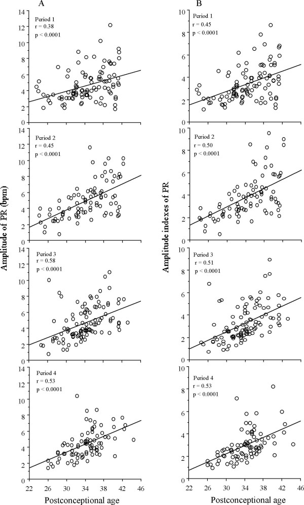
Results: Circadian cycles were observed among 23.8% neonates in HR, 20% in PR, 27.8% in RR and 16% in SpO2 in 0-3 days of PNA. Percentages of circadian cycles were the highest (40%) at < 28 wks of gestational age (GA), decreasing with GA, and the lowest (14.3%) at > or = 37 wks GA within 3 days of PNA in PR and were decreased in the later PNA. An increase of the amplitude with GA was observed in PR, and significant group differences were present in all periods. Amplitudes and amplitude indexes were positively correlated with postconceptional age (PCA) in PR (p < 0.001). Among clinical parameters, oxygen administration showed significant association (p < 0.05) with circadian rhythms of PR in the first 3 days of life.
Conclusion: Whereas circadian rhythmicity in neonates may result from maternal influence, the increase of amplitude indexes in PR with PCA may be related to physiological maturity. Further studies are needed to elucidate the effect of oxygenation on physiological rhythmicity in neonates.
Figures
References
-
- Hammarlund K, Stromberg B, Sedin G. Heat loss from the skin of preterm and fullterm newborn infants during the first weeks after birth. Biol Neonate. 1986;50:1–10. - PubMed
-
- Bauer K, Versmold H. Postnatal weight loss in preterm neonates less than 1,500 g is due to isotonic dehydration of the extracellular volume. Acta Paediatr Scand Suppl. 1989;360:37–42. - PubMed
LinkOut - more resources
Full Text Sources
Research Materials
