

Divergent projections of catecholaminergic neurons in the nucleus of the solitary tract to limbic forebrain and medullary autonomic brain regions
- PMID: 16962080
- PMCID: PMC1876790
- DOI: 10.1016/j.brainres.2006.08.051
Divergent projections of catecholaminergic neurons in the nucleus of the solitary tract to limbic forebrain and medullary autonomic brain regions
Abstract
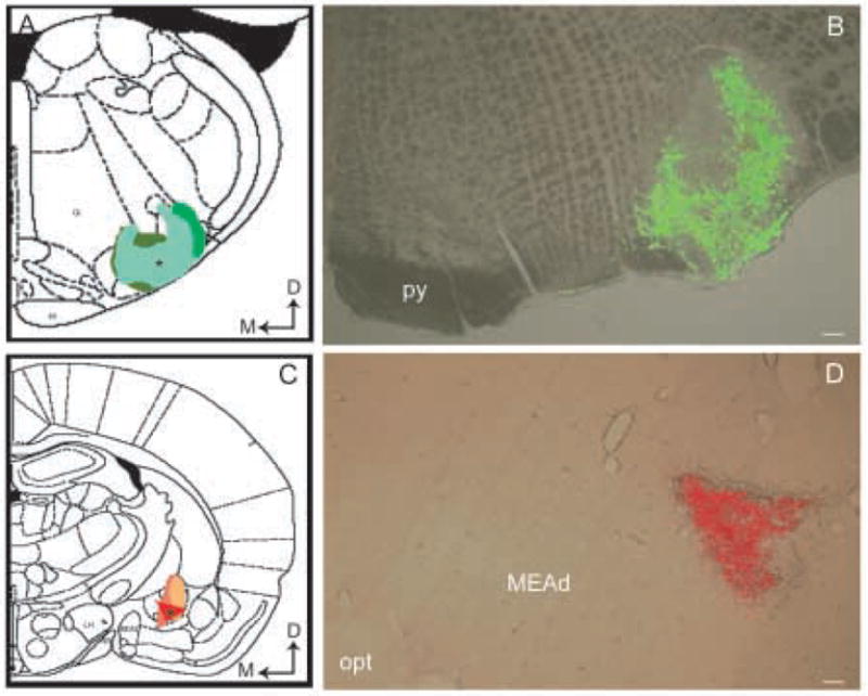
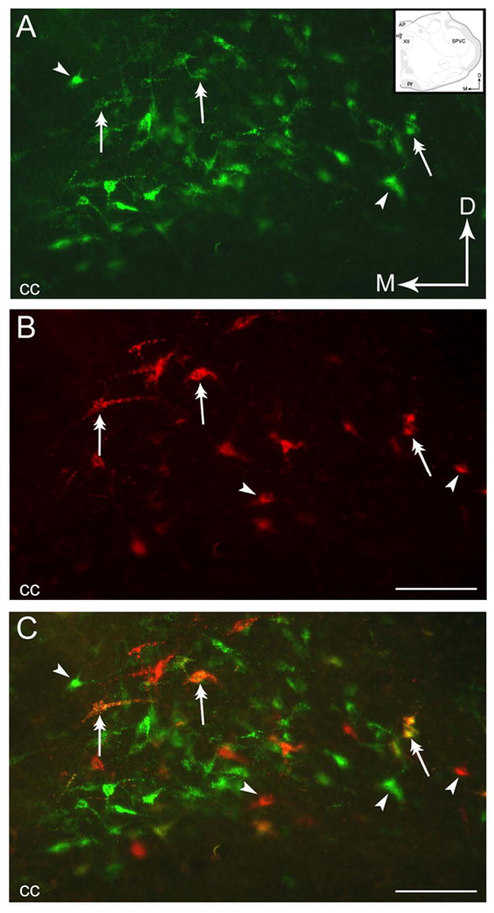
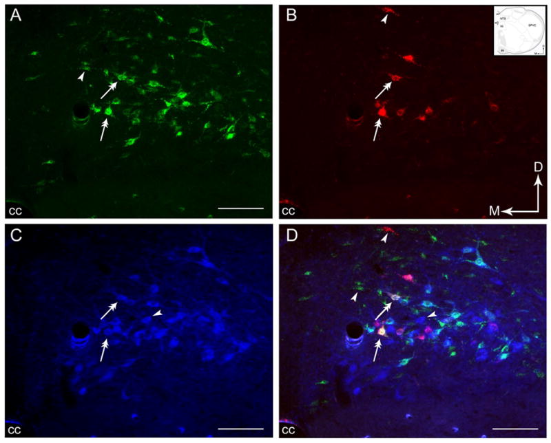
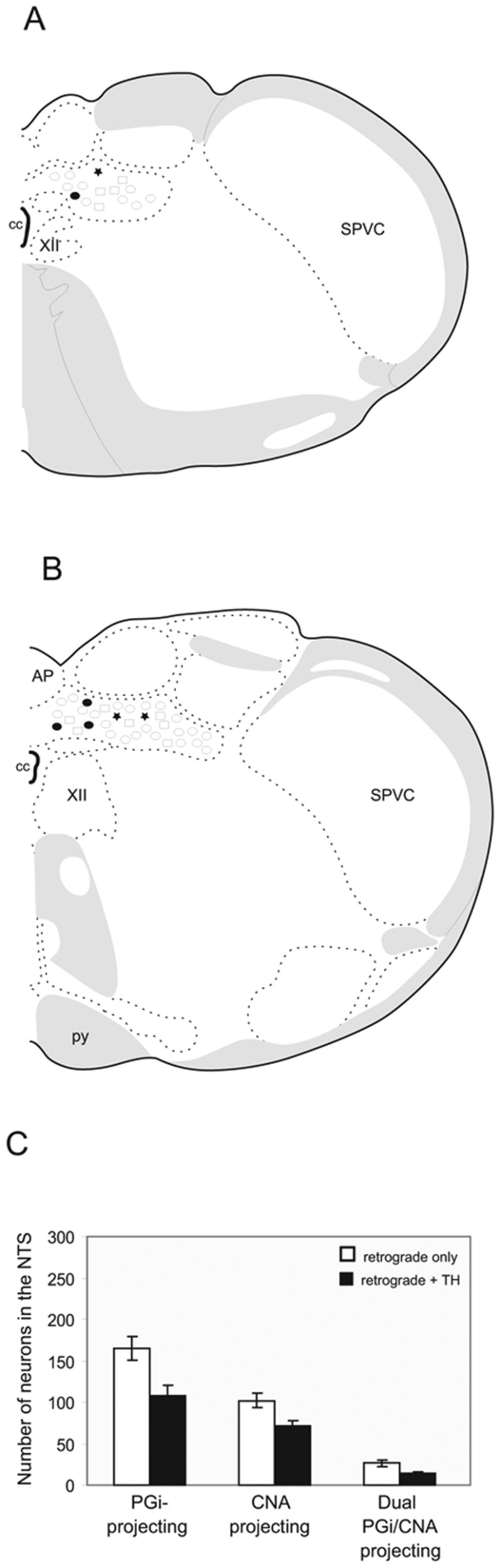
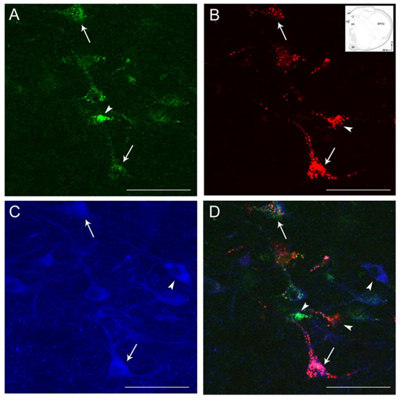
The nucleus of the solitary tract (NTS) is a critical structure involved in coordinating autonomic and visceral activities. Previous independent studies have demonstrated efferent projections from the NTS to the nucleus paragigantocellularis (PGi) and the central nucleus of the amygdala (CNA) in rat brain. To further characterize the neural circuitry originating from the NTS with postsynaptic targets in the amygdala and medullary autonomic targets, distinct green or red fluorescent latex microspheres were injected into the PGi and the CNA, respectively, of the same rat. Thirty-micron thick tissue sections through the lower brainstem and forebrain were collected. Every fourth section through the NTS region was processed for immunocytochemical detection of tyrosine hydroxylase (TH), a marker of catecholaminergic neurons. Retrogradely labeled neurons from the PGi or CNA were distributed throughout the rostro-caudal segments of the NTS. However, the majority of neurons containing both retrograde tracers were distributed within the caudal third of the NTS. Cell counts revealed that approximately 27% of neurons projecting to the CNA in the NTS sent collateralized projections to the PGi while approximately 16% of neurons projecting to the PGi sent collateralized projections to the CNA. Interestingly, more than half of the PGi and CNA-projecting neurons in the NTS expressed TH immunoreactivity. These data indicate that catecholaminergic neurons in the NTS are poised to simultaneously coordinate activities in limbic and medullary autonomic brain regions.
Figures
References
-
- Aicher SA, et al. Monosynaptic projections from the nucleus tractus solitarii to C1 adrenergic neurons in the rostral ventrolateral medulla: comparison with input from the caudal ventrolateral medulla. J Comp Neurol. 1996;373:62–75. - PubMed
-
- Allen GV, Cechetto DF. Functional and anatomical organization of cardiovascular pressor and depressor sites in the lateral hypothalamic area: I. Descending projections. J Comp Neurol. 1992;315:313–332. - PubMed
-
- Altschuler SM, et al. Viscerotopic representation of the upper alimentary tract in the rat: sensory ganglia and nuclei of the solitary and spinal trigeminal tracts. J Comp Neurol. 1989;283:248–268. - PubMed
-
- Buller KM, et al. NTS catecholamine cell recruitment by hemorrhage and hypoxia. Neuroreport. 1999;10:3853–3856. - PubMed
-
- Chan RK, Sawchenko PE. Hemodynamic regulation of tyrosine hydroxylase messenger RNA in medullary catecholamine neurons: a c-fos-guided hybridization histochemical study. Neuroscience. 1995;66:377–390. - PubMed
Publication types
MeSH terms
Substances
Grants and funding
LinkOut - more resources
Full Text Sources
