Comparative Study
. 2006 Oct 13;363(1):63-74.
doi: 10.1016/j.jmb.2006.07.021. Epub 2006 Jul 15.

Sites of interaction of a precursor polypeptide on the export chaperone SecB mapped by site-directed spin labeling

Affiliations
Comparative Study

Sites of interaction of a precursor polypeptide on the export chaperone SecB mapped by site-directed spin labeling

Jennine M Crane et al. J Mol Biol. .

Abstract

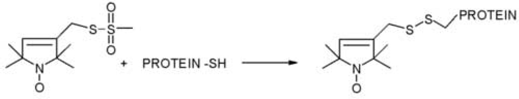
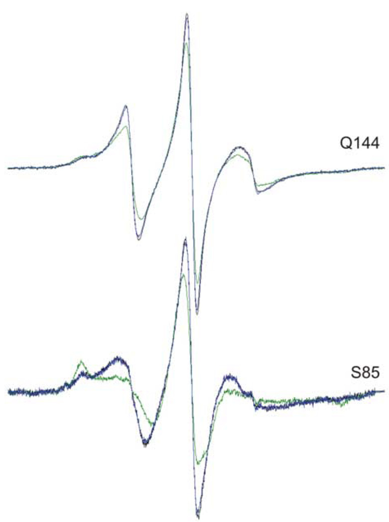
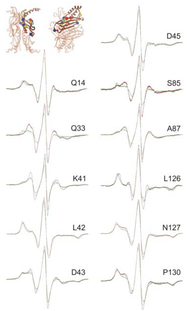
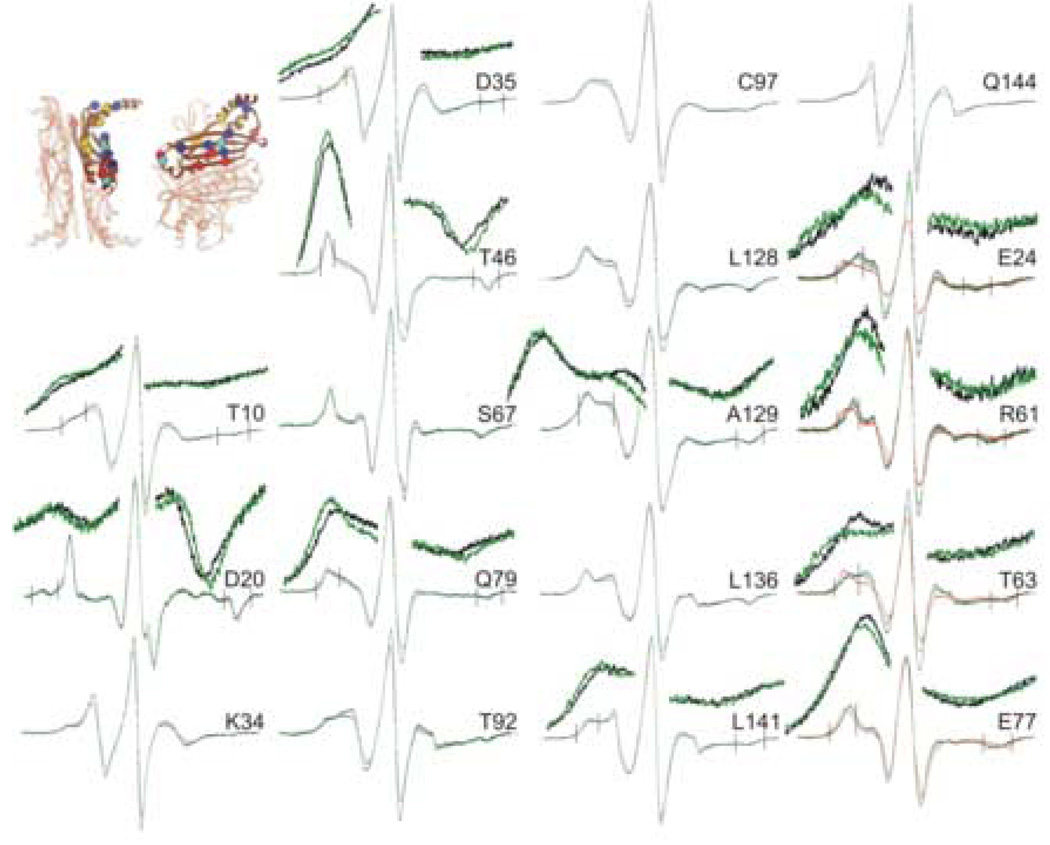
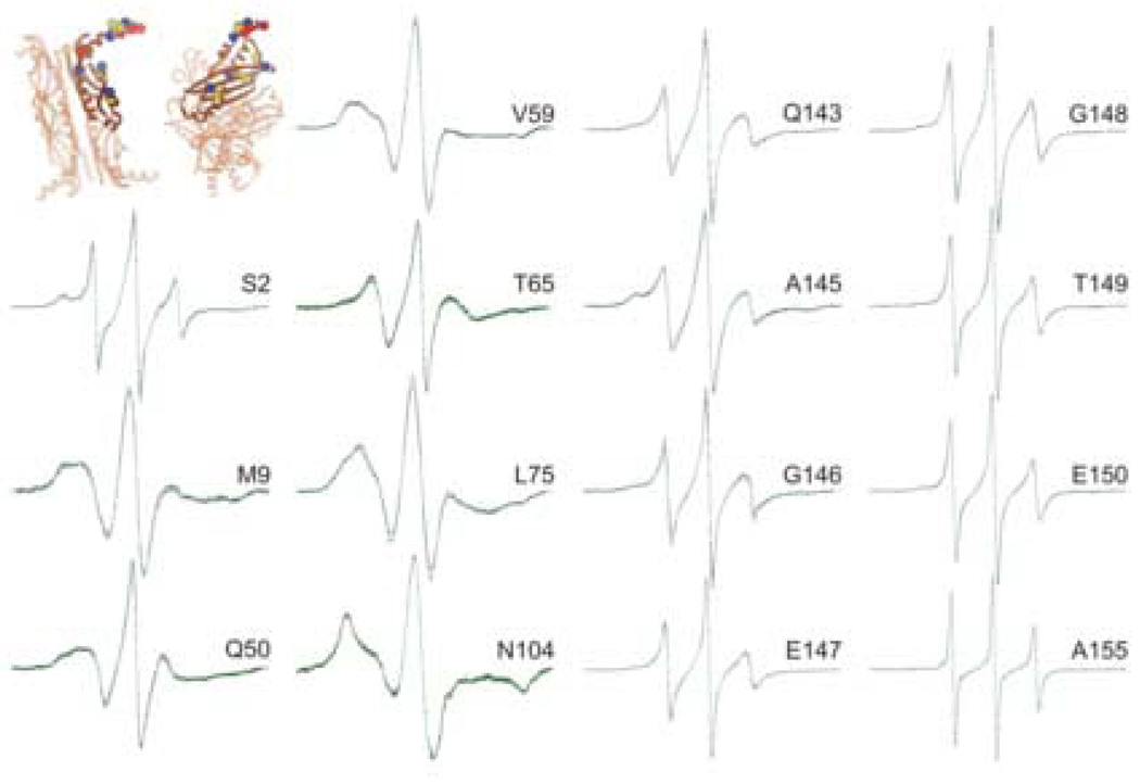
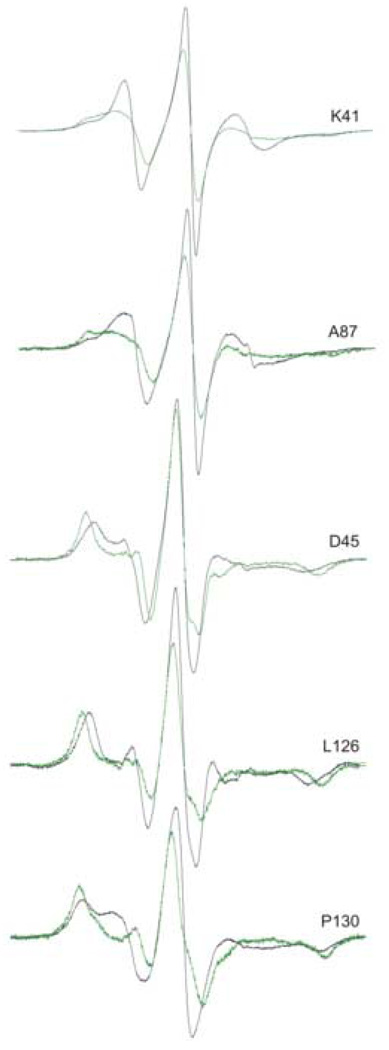
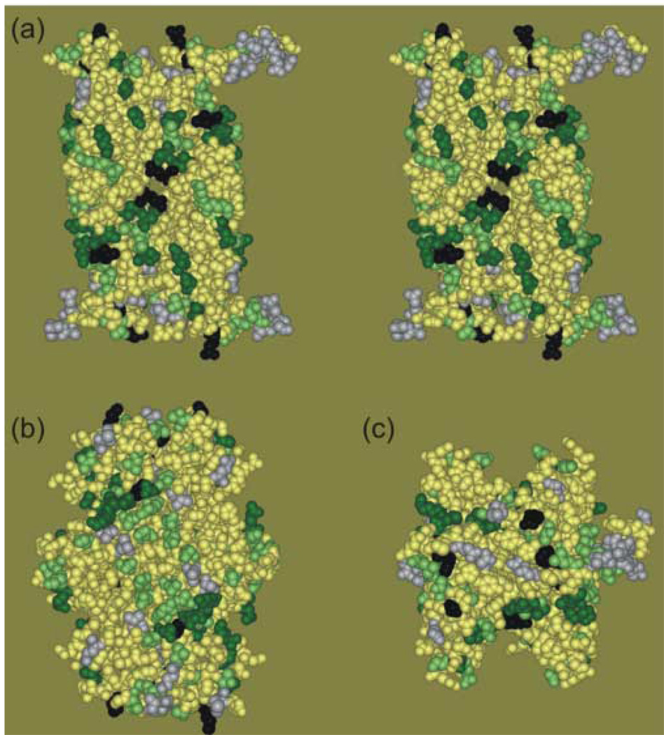
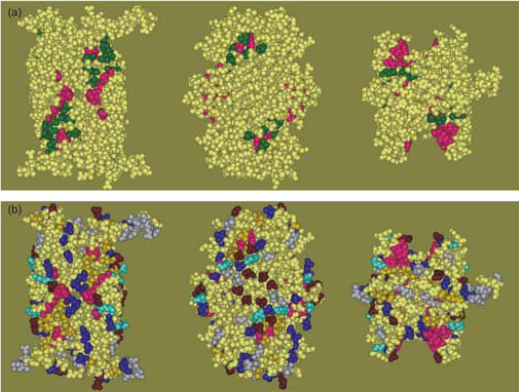
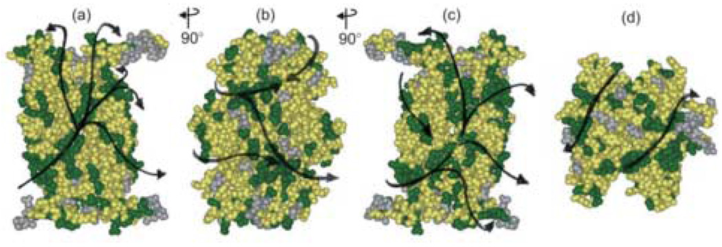
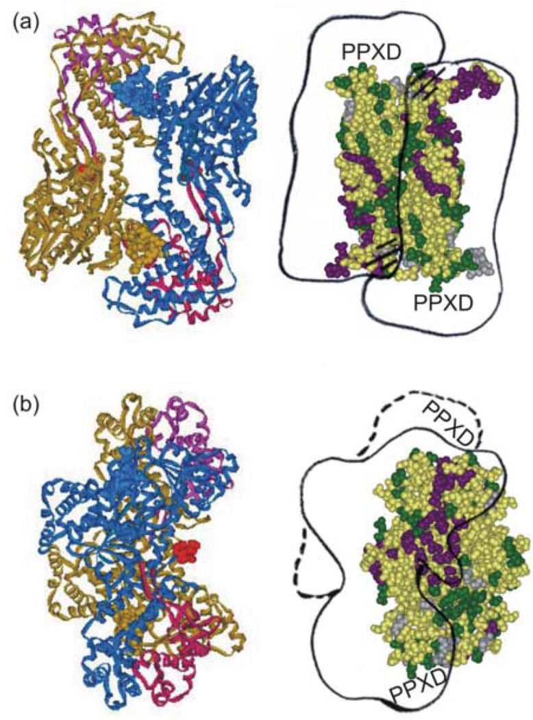
Export of protein into the periplasm of Escherichia coli via the general secretory system requires that the transported polypeptides be devoid of stably folded tertiary structure. Capture of the precursor polypeptides before they fold is achieved by the promiscuous binding to the chaperone SecB. SecB delivers its ligand to export sites through its specific binding to SecA, a peripheral component of the membrane translocon. At the translocon the ligand is passed from SecB to SecA and subsequently through the SecYEG channel. We have previously used site-directed spin labeling and electron paramagnetic resonance spectroscopy to establish a docking model between SecB and SecA. Here we report use of the same strategy to map the pathway of a physiologic ligand, the unfolded form of precursor galactose-binding protein, on SecB. Our set of SecB variants each containing a single cysteine, which was used in the previous study, has been expanded to 48 residues, which cover 49% of the surface of SecB. The residues on SecB involved in contacts were identified as those that, upon addition of the unfolded polypeptide ligand, showed changes in spectral line shape consistent with restricted motion of the nitroxide. We conclude that the bound precursor makes contact with a large portion of the surface of the small chaperone. The sites on SecB that interact with the ligand are compared with the previously identified sites that interact with SecA and a model for transfer of the ligand is discussed.


Figures

Figure 1
Figure 1
Measures of mobility. The peak to peak width of the central resonance line (ΔHpp) is measured as indicated and is equivalent to the peak width at half-height of an absorbance spectrum. The spectral breadth (2A'zz) is the distance between the outermost hyperfine extrema. The spectrum used to illustrate these parameters is that of L67R1.
Figure 2
Figure 2
The methanethiosulfonate spin label and the side chain (R1) it generates.
Figure 3
Figure 3
Reversal of complex formation between precursor galactose-binding protein and SecB. Black traces are free SecB, green traces are SecB and galactose-binding protein, blue traces are SecB and galactose-binding protein after addition of 3 mM CaCl2 and incubation at room temperature for 30 minutes before acquiring the spectrum at 6°C.
Figure 4
Figure 4
Positions on SecB that show a high degree of constraint with precursor galactose-binding protein bound. The black traces are spectra of spin-labeled SecB alone, the red are spectra of an equimolar mixture of SecA and spin-labeled SecB and the green are an equimolar mixture of spin-labeled SecB and unfolded precursor galactose-binding protein. The positions of the residues examined are shown on the structure of SecB as spheres at the site of the α-carbon atom. The color indicates the nature of the original residue: gold, hydrophobic; blue, polar; bright blue, negative charge; red, positive charge. The structure in this and all subsequent structures is that generated by threading the E. coli sequence through the H. influenzae structure because the C-terminal residues are resolved. The PDB code for SecB is 1FX3.
Figure 5
Figure 5
Positions on SecB that show significant constraint. The black traces are spectra of spin-labeled SecB alone, the red are spectra of an equimolar mixture of SecA and spin-labeled SecB and the green are an equimolar mixture of spin-labeled SecB and unfolded precursor galactose-binding protein. The positions of the residues examined are shown on the structure of SecB as spheres at the site of the α-carbon atom. The key to the colors is described in Figure 4. The insets show the region of the extrema, indicated by the vertical lines, magnified by a factor of four for both the intensity and the field.
Figure 6
Figure 6
Positions on SecB that show no significant change when precursor galactose-binding protein binds. The black traces are spectra of spin-labeled SecB alone, the red are spectra of an equimolar mixture of SecA and spin-labeled SecB and the green are an equimolar mixture of spin-labeled SecB and unfolded precursor galactose-binding protein. The positions of the residues examined are shown on the structure of SecB as spheres at the site of the α-carbon atom. The key to the colors is described in Figure 4.
Figure 7
Figure 7
Titration of spectra to reveal the state of nitroxide in contact sites. The spectra shown were generated by taking the difference between the normalized spectrum of the complex and the normalized spectrum of SecB reduced as described in the text. The amount of the reduction of the SecB spectrum was 41% for K41R1, 39% for A87R1, 31% for D45R1, 43% for L126R1 and 52% for P130R1. The titrated spectra were normalized to have the same total spin as the spectra of the complexes.
Figure 8
Figure 8
Contact sites for precursor galactose-binding protein on SecB. The residues analyzed by site-directed spin labeling are shown on the structure as CPK models of the residue that was substituted by cysteine and derivatized with the spin label. The positions that showed a high degree of change in spectral line shape when precursor galactose-binding protein was added are shown in dark green, those that showed a significant change in blue green and those that showed no change in gray. The residues in black are assigned to the binding site because derivatization inactivated SecB for binding precursor. Panel (a) is a stereo view with the dimer interface facing forward. Panel (b) is related to panel (a) by a 90° rotation about the vertical axis to the right. This view shows the flat β-sheet on the side of the tetramer. Panel (c) is related to panel (a) by a 90° rotation about the horizontal axis toward the viewer. This view shows the top of the tetramer.
Figure 9
Figure 9
Panel (a) Binding site previously proposed based on the crystal structure. The structure on the left is a view of the dimer interface. The view in the center was achieved by a 90° rotation about the vertical axis to show the flat -sheet on the side of the tetramer. The view on the right is related to that on the left by a 90° rotation about the horizontal axis toward the viewer. Panel (b) Contact sites identified in this study colored to indicate the nature of the residue. The color scheme is described in the text. The orientation of the structure is as described in panel (a).
Figure 10
Figure 10
Possible pathways around SecB. All contact sites identified in this study are shown in green. The arrows indicate possible routes around the chaperone. (a), (b) and (c) are each related to the previous structure by a 90° rotation about the vertical axis to the left. (d) is related to (a) by a 90° rotation about the horizontal axis toward the viewer. (a) front view of the deep channel; the interface of the dimers that form the tetramer is aligned with the vertical axis. (b) flat 8-stranded β-sheet that is the side of the tetramer. (c) channel of the dimer interface at the face opposite that shown in (a). (d) end view of the tetramer.
Figure 11
Figure 11
Simultaneous binding of precursor galactose-binding protein and SecA. Panel (a) left side, structure of the SecA dimer oriented so that the extreme C-termini, which are not resolved, and the PPXD (magenta and pink) are on the lower surface. Right side, an outline of the SecA dimer is docked across SecB as described in the text. The hatched lines indicate the binding sites on SecA that interact with the flexible C-terminal regions of SecB (shown in structure as helical). Panel (b) structures shown are related to those in panel (a) by a 90° rotation about the vertical axis to the left. The PDB code for SecA is 1M74.

References

    1. Berks BC, Sargent F, Palmer T. The Tat protein export pathway. Mol. Microbiol. 2000;35:260–274. - PubMed
    1. Economou A. Bacterial secretome: the assembly manual and operating instructions. Molecular Membrane Biology. 2002;19:159–169. - PubMed
    1. Randall LL, Hardy SJS. High selectivity with low specificity: how SecB has solved the paradox of chaperone binding. Trends Biochem. Sci. 1995;20:65–69. - PubMed
    1. Crane JM, Mao C, Lilly AA, Smith VF, Suo Y, Hubbell WL, Randall LL. Mapping of the docking of SecA onto the chaperone SecB by site-directed spin labeling: insight into the mechanism of ligand transfer during protein export. J. Mol. Biol. 2005;353:295–307. - PubMed
    1. Topping TB, Randall LL. Determination of the binding frame within a physiological ligand for the chaperone SecB. Protein Sci. 1994;3:730–736. - PMC - PubMed

Publication types

MeSH terms

LinkOut - more resources