

Emergence of a spermine-sensitive, non-inactivating conductance in mature hippocampal CA1 pyramidal neurons upon reduction of extracellular Ca2+: dependence on intracellular Mg2+ and ATP
- PMID: 16962211
- PMCID: PMC1853290
- DOI: 10.1016/j.neuint.2006.07.013
Emergence of a spermine-sensitive, non-inactivating conductance in mature hippocampal CA1 pyramidal neurons upon reduction of extracellular Ca2+: dependence on intracellular Mg2+ and ATP
Abstract
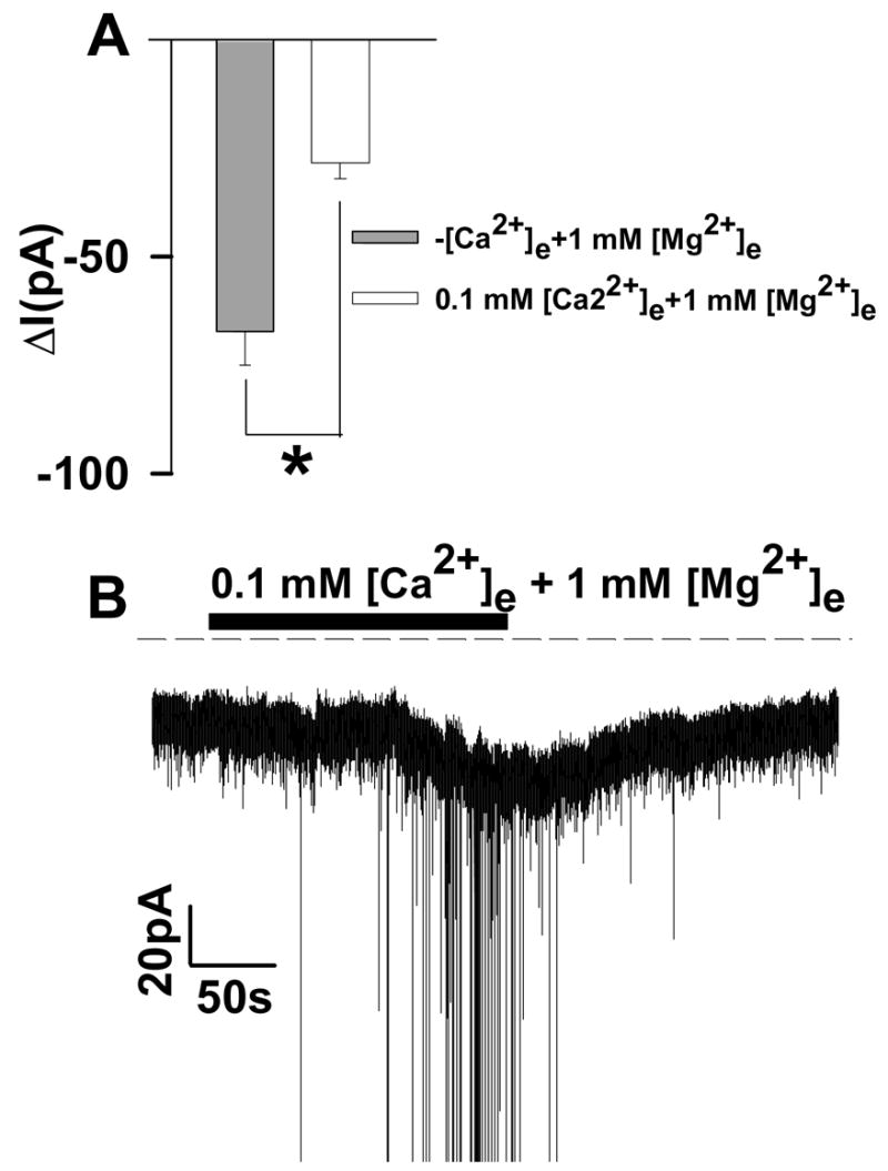
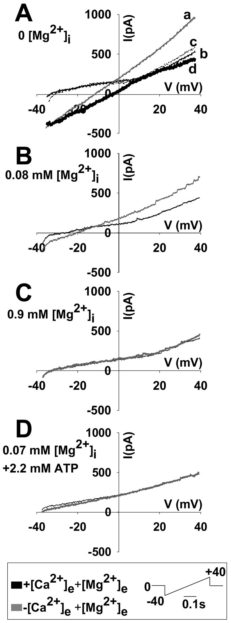
Large and protracted elevations of intracellular [Ca(2+)] and [Na(+)] play a crucial role in neuronal injury in ischemic conditions. In addition to excessive glutamate receptor activation, other ion channels may contribute to disruption of intracellular ionic homeostasis. During episodes of ischemia, extracellular [Ca(2+)] falls significantly. Here we report the emergence of an inward current in hippocampal CA1 pyramidal neurons in acute brain slices from adult mice upon reduction/removal of [Ca(2+)](e). The magnitude of the current was 100-300pA at -65mV holding potential, depending on intracellular constituents. The current was accompanied by intense neuronal discharge, observed in both whole-cell and cell-attached patch configurations. Sustained currents and increased neuronal firing rates were both reversed by restoration of physiological levels of [Ca(2+)](e), or by application of spermine (1mM). The amplitudes of the sustained currents were strongly reduced by raising intracellular [Mg(2+)], but not by extracellular [Mg(2+)] increases. Elevated intracellular ATP also reduced the current. This conductance is similar in several respects to the "calcium-sensing, non-selective cation current" (csNSC), previously described in cultured mouse hippocampal neurons of embryonic origin. The dependence on intracellular [ATP] and [Mg(2+)] shown here, suggests a possible role for this current in disruption of ionic homeostasis during metabolic stress that accompanies excessive neuronal stimulation.
Figures


Similar articles
-
Acidosis decreases low Ca(2+)-induced neuronal excitation by inhibiting the activity of calcium-sensing cation channels in cultured mouse hippocampal neurons.J Physiol. 2003 Jul 15;550(Pt 2):385-99. doi: 10.1113/jphysiol.2003.043091. Epub 2003 May 30. J Physiol. 2003. PMID: 12777448 Free PMC article.
-
Effect of lamotrigine on the Ca(2+)-sensing cation current in cultured hippocampal neurons.J Neurophysiol. 2001 Nov;86(5):2520-6. doi: 10.1152/jn.2001.86.5.2520. J Neurophysiol. 2001. PMID: 11698539
-
Blockade of Ca2+-permeable AMPA/kainate channels decreases oxygen-glucose deprivation-induced Zn2+ accumulation and neuronal loss in hippocampal pyramidal neurons.J Neurosci. 2002 Feb 15;22(4):1273-9. doi: 10.1523/JNEUROSCI.22-04-01273.2002. J Neurosci. 2002. PMID: 11850455 Free PMC article.
-
Spermine blocks synaptic transmission mediated by Ca(2+)-permeable AMPA receptors.Neuroreport. 1996 Feb 29;7(3):689-92. doi: 10.1097/00001756-199602290-00002. Neuroreport. 1996. PMID: 8733722
-
Effects of extracellular pH on voltage-gated Na+, K+ and Ca2+ currents in isolated rat CA1 neurons.J Physiol. 1996 Jun 15;493 ( Pt 3)(Pt 3):719-32. doi: 10.1113/jphysiol.1996.sp021417. J Physiol. 1996. PMID: 8799894 Free PMC article.
Cited by
-
Zn²+ chelation improves recovery by delaying spreading depression-like events.Neuroreport. 2010 Nov 17;21(16):1060-4. doi: 10.1097/WNR.0b013e32833fd42c. Neuroreport. 2010. PMID: 20856149 Free PMC article.
-
Non-canonical glutamate signaling in a genetic model of migraine with aura.Neuron. 2021 Feb 17;109(4):611-628.e8. doi: 10.1016/j.neuron.2020.11.018. Epub 2020 Dec 14. Neuron. 2021. PMID: 33321071 Free PMC article.
-
Localized loss of Ca2+ homeostasis in neuronal dendrites is a downstream consequence of metabolic compromise during extended NMDA exposures.J Neurosci. 2008 May 7;28(19):5029-39. doi: 10.1523/JNEUROSCI.5069-07.2008. J Neurosci. 2008. PMID: 18463256 Free PMC article.
-
Oxidative Stress in the Hypothalamus: the Importance of Calcium Signaling and Mitochondrial ROS in Body Weight Regulation.Curr Neuropharmacol. 2012 Dec;10(4):344-53. doi: 10.2174/157015912804143496. Curr Neuropharmacol. 2012. PMID: 23730258 Free PMC article.
-
Tonic inhibition of TRPV3 by Mg2+ in mouse epidermal keratinocytes.J Invest Dermatol. 2012 Sep;132(9):2158-65. doi: 10.1038/jid.2012.144. Epub 2012 May 24. J Invest Dermatol. 2012. PMID: 22622423 Free PMC article.
References
-
- Aarts M, Iihara K, Wei WL, Xiong ZG, Arundine M, Cerwinski W, MacDonald JF, Tymianski M. A key role for TRPM7 channels in anoxic neuronal death. Cell. 2003;115:863–877. - PubMed
-
- Aitken PG, Breese GR, Dudek FF, Edwards F, Espanol MT, Larkman PM, Lipton P, Newman GC, Nowak TS, Jr, Panizzon KL. Preparative methods for brain slices: a discussion. J Neurosci Methods. 1995;59:139–149. - PubMed
-
- Arundine M, Tymianski M. Molecular mechanisms of calcium-dependent neurodegeneration in excitotoxicity. Cell Calcium. 2003;34:325–337. - PubMed
Publication types
MeSH terms
Substances
Grants and funding
LinkOut - more resources
Full Text Sources
Miscellaneous
