Review
. 2006 Sep;25(5):490-513.
doi: 10.1016/j.preteyeres.2006.07.003. Epub 2006 Sep 7.

Oxidative stress in glaucomatous neurodegeneration: mechanisms and consequences

Affiliations
Review

Oxidative stress in glaucomatous neurodegeneration: mechanisms and consequences

Gülgün Tezel. Prog Retin Eye Res. 2006 Sep.

Abstract

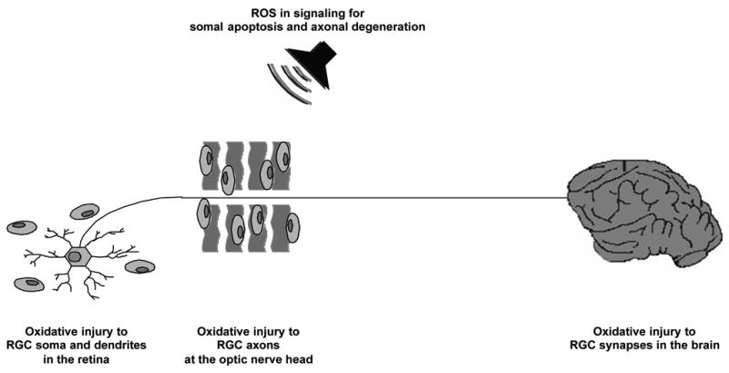
Reactive oxygen species (ROS) are generated as by-products of cellular metabolism, primarily in the mitochondria. Although ROS are essential participants in cell signaling and regulation, when their cellular production overwhelms the intrinsic antioxidant capacity, damage to cellular macromolecules such as DNA, proteins, and lipids ensues. Such a state of "oxidative stress" is thought to contribute to the pathogenesis of a number of neurodegenerative diseases. Growing evidence supports the involvement of oxidative stress as a common component of glaucomatous neurodegeneration in different subcellular compartments of retinal ganglion cells (RGCs). Besides the evidence of direct cytotoxic consequences leading to RGC death, it also seems highly possible that ROS are involved in signaling RGC death by acting as a second messenger and/or modulating protein function by redox modifications of downstream effectors through enzymatic oxidation of specific amino acid residues. Different studies provide cumulating evidence, which supports the association of ROS with different aspects of the neurodegenerative process. Oxidative protein modifications during glaucomatous neurodegeneration increase neuronal susceptibility to damage and also lead to glial dysfunction. Oxidative stress-induced dysfunction of glial cells may contribute to spreading neuronal damage by secondary degeneration. Oxidative stress also promotes the accumulation of advanced glycation end products in glaucomatous tissues. In addition, oxidative stress takes part in the activation of immune response during glaucomatous neurodegeneration, as ROS stimulate the antigen presenting ability of glial cells and also function as co-stimulatory molecules during antigen presentation. By discussing current evidence, this review provides a broad perspective on cellular mechanisms and potential consequences of oxidative stress in glaucoma.


Figures

Figure 1
Figure 1
Multiple pathogenic mechanisms have been proposed for glaucomatous neurodegeneration. Most of these mechanisms appear to be associated with a common pathway of oxidative injury. Although the initial site of glaucomatous injury is unclear, RGC survival and axon health are dependent on each other; and primary injury to any of these subcellular compartments subsequently affects the others.
Figure 2
Figure 2
RGC death induced by glaucomatous stimuli involves receptor-mediated caspase activation, and caspase-dependent and -independent components of the mitochondrial cell death pathway. Amplified ROS generation and oxidative damage are associated with both receptor-mediated and mitochondria-mediated pathways of RGC death. A critical balance between a variety of intracellular signaling pathways linked to cell survival or cell death determines whether a RGC dies or survives the glaucomatous stress (IOP, intraocular pressure; ROS, reactive oxygen species; bcl-xL and bax, anti-apoptotic and pro-apoptotic members of the bcl-2 family of proteins, respectively; aif and c, mitochondrial cell death mediators, apoptosis inducing factor and cytochrome c, respectively; TNF-α, tumor necrosis factor-alpha; TNF-R1, TNF death receptor; NO, nitric oxide; NF-κB, nuclear factor-kappaB, transcription factor; IκB, NF-κB inhibiting protein; JNK and ERK, mitogen-activated protein kinases, c-jun N-terminal kinase and extracellular signal-regulated kinase, respectively).
Figure 3
Figure 3
In addition to neurodegenerative injury induced by intracellular ROS, ROS released from stressed cells into the extracellular milieu may also facilitate secondary degeneration of RGCs. ROS released by neighboring cells may be directly cytotoxic to primarily undamaged RGCs. Alternatively, various consequences of oxidative stress-induced dysfunction of supportive glia may take part in spreading neuronal damage by secondary degeneration of RGCs. By leading to glial dysfunction, oxidative stress-induced glial activation, glial protein oxidation, and AGE/RAGE signaling may all contribute to decreased glial support of RGCs. By stimulating the antigen presenting ability of glial cells, ROS may also be involved in aberrant activation of the immune system, thereby facilitating the progression of glaucomatous neurodegeneration.
Figure 4
Figure 4
Glaucomatous neurodegeneration evidently exhibits widespread damage through a set of compartmentalized subcellular processes, which eventually involves RGC soma and dendrites in the retina, axons in the optic nerve, and synapses in the brain. Increasing evidence supports that widespread neurodegenerative cascades in glaucoma likely involve a common oxidative component. In addition to cytotoxic events induced by amplified ROS generation, ROS can also function as signaling molecules during glaucomatous neurodegeneration in different subcellular compartments of RGCs. ROS can act as a second messenger and/or modulate protein function by redox modifications of downstream effectors.
Figure 5
Figure 5
Proteomic analysis was performed to determine whether retinal proteins are oxidatively modified during glaucomatous neurodegeneration in ocular hypertensive eyes following hypertonic saline injections into episcleral veins. Protein expression was determined by two dimensional (2D)-polyacrylamide gel electrophoresis of equally loaded protein samples obtained from ocular hypertensive and control retinas (PI, isoelectric point). Protein oxidation was determined by identifying the retinal proteins containing carbonyl groups using 2D-oxyblot analysis. Results of this in vivo study revealed that the protein modification by ROS occurs to a greater extent in ocular hypertensive eyes compared with the controls. Comparison of 2D-oxyblots with Coomassie Blue-stained 2D-gels showed that approximately 60 protein spots (out of over 400 spots) obtained using retinal protein lysates from ocular hypertensive retinas exhibited protein carbonyl immunoreactivity, which reflects oxidatively modified proteins. Following normalization of spots to their protein content measured by the intensity of Coomassie Blue staining, a significant increase was detected in carbonyl immunoreactivity of individual protein spots obtained using retinal protein lysates from ocular hypertensive eyes compared with the controls. The oxidized proteins identified through mass spectrometry included glyceraldehyde-3-phosphate dehydrogenase (GAPDH), a glycolytic enzyme; HSP72, a stress protein; and glutamine synthetase, an excitotoxicity-related protein (Modified with permission from (Tezel et al., 2005).

Similar articles

Cited by

References

    1. Abu-Amero KK, Morales J, Bosley TM. Mitochondrial abnormalities in patients with primary open-angle glaucoma. Invest Ophthalmol Vis Sci. 2006;47:2533–2541. - PubMed
    1. Agapova OA, Ricard CS, Salvador-Silva M, Hernandez MR. Expression of matrix metalloproteinases and tissue inhibitors of metalloproteinases in human optic nerve head astrocytes. Glia. 2001;33:205–216. - PubMed
    1. Andersen JK. Oxidative stress in neurodegeneration: cause or consequence? Nat Med. 2004;10:S18–25. Suppl. - PubMed
    1. Anderson DR, Hendrickson A. Effect of intraocular pressure on rapid axoplasmic transport in monkey optic nerve. Invest Ophthalmol. 1974;13:771–783. - PubMed
    1. Araie M. Pattern of visual field defects in normal-tension and high-tension glaucoma. Curr Opin Ophthalmol. 1995;6:36–45. - PubMed

Publication types