. 2007 Jan 1;301(1):178-91.
doi: 10.1016/j.ydbio.2006.08.011. Epub 2006 Aug 10.

Tissue remodeling during maturation of the Drosophila wing

Affiliations

Tissue remodeling during maturation of the Drosophila wing

John A Kiger Jr et al. Dev Biol. .

Abstract

The final step in morphogenesis of the adult fly is wing maturation, a process not well understood at the cellular level due to the impermeable and refractive nature of cuticle synthesized some 30 h prior to eclosion from the pupal case. Advances in GFP technology now make it possible to visualize cells using fluorescence after cuticle synthesis is complete. We find that, between eclosion and wing expansion, the epithelia within the folded wing begin to delaminate from the cuticle and that delamination is complete when the wing has fully expanded. After expansion, epithelial cells lose contact with each other, adherens junctions are disrupted, and nuclei become pycnotic. The cells then change shape, elongate, and migrate from the wing into the thorax. During wing maturation, the Timp gene product, tissue inhibitor of metalloproteinases, and probably other components of an extracellular matrix are expressed that bond the dorsal and ventral cuticular surfaces of the wing following migration of the cells. These steps are dissected using the batone and Timp genes and ectopic expression of alphaPS integrin, inhibitors of Armadillo/beta-catenin nuclear activity and baculovirus caspase inhibitor p35. We conclude that an epithelial-mesenchymal transition is responsible for epithelial delamination and dissolution.


Figures
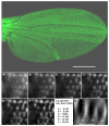
Figure 1
Figure 1
Top. Newly open wing of a fly of genotype Gal4-30A UAS-GFP. Bar = 400 μm. Bottom (A–F). Serial projections in the XY plane (viewed up the Z axis) of optical section stacks of increasing depth, prepared by epifluorescence deconvolution microscopy, of a newly open wing of the same genotype. The thickness of each stack is indicated. (G) An optical section stack rotated by 90° to make the Z axis vertical and viewed along the XY plane to present an edge-on view of this section of the wing (apical surface of the epithelium is down, basal up). Arrows indicate the positions of adjacent wing hairs. Bar = 15 μm.
Figure 2
Figure 2
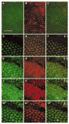
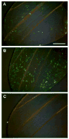
Labeling of wing cells by ywing-GFP (green) and Gal-4-30A driven UAS-AUG-DsRed (red). (A–C) Cells in a newly open adult wing viewed using epifluorescence: green (A); red (B); merger (C). Bar = 50 μm. (D–O) Cells in a pupal wing at stage P11 viewed in a series of confocal optical sections (Z); green, red and merger ordered as for (A–C). The pupal wing hairs autofluoresce in the red channel (E). The boundary between dorsal and ventral epithelia is imaged in Z5.
Figure 3
Figure 3
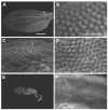
Delamination of epithelial cells from the wing cuticle in normal and batone mutant flies of genotype apterous-Gal4 UAS-GFP/+. (A) Wing (10 minutes after opening) of a batone/+ female. (B) Magnified view of A. Note that the wing hairs are centered between areas of maximum fluorescence. (C) Magnified view of one unopened wing of a newly eclosed fly. Note the lighter cuticular ridges outlining the epithelial cells and the uniform distribution of fluorescence as compared to B. (D) Magnified view of another region of an unopened wing of the same newly eclosed fly. Note that the wing hairs are centered between areas of maximum fluorescence in this portion of the wing, as in B. (E) Wing of a batone/Y male aged 1–2 hr after eclosion has not opened. (F) Magnified view of a wing of a batone/Y male aged 21–28 hr after eclosion is similar to that in C. The wing has not opened, and no delamination is apparent. In A and E, bar = 400 μm; in B, C, D and F, bar = 50 μm.
Figure 4
Figure 4
Epithelial cells disappear from the wing following delamination from the cuticle. Flies of genotype Gal4-30A UAS-lacZ/UAS-GFP were allowed to open their wings fully and then, at recorded times, wings were removed, fixed, and stained for LacZ activity as described in Materials and Methods. The minutes between wing opening and fixation are indicated for each wing. The bar = 400 μm.
Figure 5
Figure 5
Epithelial cells break contacts with each other, change shape, and change position following opening of the wing. (A) Wing from a fly of genotype apterous-Gal4/UAS-GFP. Note the close contacts of the cells. (B) The same wing viewed 124 minutes later. (C) Magnified view of cells in the central part of the wing. (D) Magnified view of cells in the transitional zone between central and distal regions that appear to be changing from round to spindle shaped. Note the fine cytoplasmic filaments extending from some of the cells. (E) Magnified view of the anterior portion of the wing in B. The directionality of the spindle shaped cells suggests streaming. In A and B, bar = 200 μm; in C, D and E, bar = 50 μm.
Figure 6
Figure 6
The effect of age on Arm-GFP fluorescence in wings. Wings are from flies of genotype y w f36a/Y; arm-GFP/+. The time at which wings were fixed following eclosion is (A) 2 minutes, (B) 40 minutes, and (C) 60 minutes. Note the DAPI-stained nuclei (false-colored magenta) in C compared to in A and B. The bar = 20 μm.
Figure 7
Figure 7
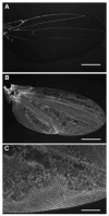
The effect of UV irradiation on epithelial cell behavior in flies of genotype apterous-Gal4/UAS-GFP. (A) Wing of a control fly that was not irradiated removed 1 hr after opening. (B) Wing of an irradiated fly removed 1 hr after opening. Although it is not obvious, the dorsal and ventral surfaces of the wing have not bonded to each other. (C) Magnified view of the wing in B. In A and B, bar = 400 μm; in C. bar = 100 μm.
Figure 8
Figure 8
The Timp gene is active in unopened wings and required for bonding of the dorsal and ventral wing surfaces. (A) A schematic of the Timp gene showing the start and stop codons, intron splice sites, 5' and 3' UTRs, and RT-PCR primer placement. (B) Gel image of RT-PCR products obtained using template nucleic acids purified from wings of newly eclosed wild-type flies. Lanes 1 and 2 show the products from the Timp primers (DKOG81/DKOG82). Lane 1 is a control reaction with no template, and lane 2 is the result with template. Expected sizes from the Timp primers are 505 bp (DNA) and 327 bp (RNA). Lanes 3 and 4 show the products from RP49 primers (DKOG52/DKOG53). Lane 3 is a control reaction with no template, and lane 4 is the result with template. Expected sizes from the RP49 primers are 320 bp (DNA) and 258 bp (RNA). (C) Wing from a fly homozygous for Timp deficiency and ywing-GFP removed approximately 22 hr after eclosion and viewed by epifluorescence. Virtually all epithelial cells have left the wing; the arrow indicates one of several small clusters of fluorescent cells, floating in hemolymph, that remain. (D) The same wing viewed in bright field, showing fluid filled blisters resulting from failure of dorsal and ventral wing surfaces to bond.
Figure 9
Figure 9
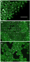
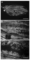
The effect of the caspase inhibitor p35 on epithelial cell behavior. Wing of a fly of genotype Gal4-30A UAS-GFP; UAS-p35 removed 18–24 hr after eclosion. (A) Bar = 400 μm. (B) Bar = 100 μm. (C) Bar = 50 μm.
Figure 10
Figure 10
Epithelial cell delamination requires both normal Armadillo/β-catenin and PS integrin functions. (A) Wing from a fly of genotype Gal4-30A/UAS-pygo3–8; ywing-GFP/+ removed >24 hr after eclosion. Many fluorescent cells remain within the wing. Note the absence of cells to the left of the vein marked with “*”; the cuticle is visible because of GFP fluorescence refracted within the cuticle. (B) Magnified view of A. The epithelial cells that have failed to delaminate from the cuticle nest within the cuticular ridges that denote the boundaries of those cells. (C) through (F) Note the failure to delaminate of significant numbers of epithelial cells of wings from flies of genotype: (C) Gal4-30A/UAS-shaggy; ywing-GFP/+; (D) Gal4-30A/UAS-Δarmadillo; ywing-GFP/+; (E) y w UAS-armadillo S10/Y; Gal4-30A/+; ywing-GFP/+ ; Inset: Gal4-30A/+; ywing-GFP/UAS-armadillo S2 ; (F) Gal4-684, UAS-αPS2m8/ywingGFP. In (A) bar = 200 μm; in (B) – (F), bar = 50 μm.
Figure 11
Figure 11
Hemese-Gal4 drives UAS-GFPnls in a small population of true hemocytes in the newly expanded wing. (A) Wing of a Hemese-Gal4 UAS-GFPnls fly. (B) Wing of a UAS-shaggy/+; Hemese-Gal4 UAS-GFPnls/+ fly. (C) Wing of a UAS-shaggy/+; Hemese-Gal4 UAS-GFPnls/+ fly open for several hours. The wing has matured normally. Bar = 200 μm.

Similar articles

Cited by

References

    1. Algeciras-Schimnich A, Barnhart BC, Peter ME. Apoptosis-independent functions of killer caspases. Curr Opin Cell Biol. 2002;14:721–726. - PubMed
    1. Ashkenas J, Muschler J, Bissell MJ. The extracellular matrix in epithelial biology: shared molecules and common themes in distant phyla. Dev Biol. 1996;180:433–444. - PMC - PubMed
    1. Axelrod JD, Matsuno K, Artavanis-Tsakonas S, Perrimon N. Interaction between Wingless and Notch signaling pathways mediated by Dishevelled. Science. 1996;271:1826–1832. - PubMed
    1. Bainbridge SP, Bownes M. Staging the metamorphosis of Drosophila melanogaster. J Embryol Exp Morph. 1981;66:57–80. - PubMed
    1. Baker JD, McNabb SL, Truman JW. The hormonal coordination of behavior and physiology at adult ecdysis in Drosophila melanogaster. J Exp Biol. 1999;202:3037–3048. - PubMed

Publication types