. 2006;34(16):4593-608.
doi: 10.1093/nar/gkl603. Epub 2006 Sep 8.

NOP132 is required for proper nucleolus localization of DEAD-box RNA helicase DDX47

Affiliations

NOP132 is required for proper nucleolus localization of DEAD-box RNA helicase DDX47

Takeshi Sekiguchi et al. Nucleic Acids Res. 2006.

Abstract

Previously, we described a novel nucleolar protein, NOP132, which interacts with the small GTP binding protein RRAG A. To elucidate the function of NOP132 in the nucleolus, we identified proteins that interact with NOP132 using mass spectrometric methods. NOP132 associated mainly with proteins involved in ribosome biogenesis and RNA metabolism, including the DEAD-box RNA helicase protein, DDX47, whose yeast homolog is Rrp3, which has roles in pre-rRNA processing. Immunoprecipitation of FLAG-tagged DDX47 co-precipitated rRNA precursors, as well as a number of proteins that are probably involved in ribosome biogenesis, implying that DDX47 plays a role in pre-rRNA processing. Introduction of NOP132 small interfering RNAs induced a ring-like localization of DDX47 in the nucleolus, suggesting that NOP132 is required for the appropriate localization of DDX47 within the nucleolus. We propose that NOP132 functions in the recruitment of pre-rRNA processing proteins, including DDX47, to the region where rRNA is transcribed within the nucleolus.


Figures
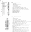
Figure 1
Figure 1
Protein components of immunoprecipitated FLAG-NOP132-, FLAG-DDX47-, and FLAG-DDX18-associated complexes. (a) Silver-stained SDS–PAGE gels of FLAG-NOP132-associated complexes immunoprecipitated with anti-FLAG from cells expressing FLAG-tagged NOP132. Lane 1, control immunoprecipitate (Mock) from untransfected 293EBNA cells; lane 2, 293EBNA cells expressing FLAG-tagged NOP132 (11% SDS–PAGE gel); lane 3, 293 cells expressing FLAG-tagged NOP132 (lysis by sonication, 10% SDS–PAGE gel)]. Molecular weight markers are indicated at the left. Because there were so many protein bands in the gels, the gel slices contained multiple proteins. The protein bands identified by mass spectrometric analysis after in-gel digestion of protein bands with protease are indicated on the right. Proteins that were also identified in the FLAG-DDX47-associated complex are underlined. (b) Silver-stained 10% SDS–PAGE gel of FLAG-DDX47-associated complexes immunoprecipitated with anti-FLAG after expression of FLAG-tagged full-length DDX47 in 293 cells. Molecular weight markers are indicated at the left. The protein bands identified by mass spectrometric analysis after in-gel digestion of protein bands with protease are indicated on the right. Proteins that were also identified in the FLAG-NOP132-associated complex are underlined. (c) Silver-stained 10% SDS–PAGE gel of FLAG-DDX18-associated complexes immunoprecipitated with anti-FLAG after expression of FLAG-tagged full-length DDX18 in 293 cells. Molecular weight markers are indicated at the left. The protein bands identified by mass spectrometric analysis after in-gel digestion of protein bands with protease are indicated on the right. Proteins that were also identified in FLAG-NOP132- and FLAG-DDX47-associated complexes are underlined and in bold, respectively.
Figure 1
Figure 1
Protein components of immunoprecipitated FLAG-NOP132-, FLAG-DDX47-, and FLAG-DDX18-associated complexes. (a) Silver-stained SDS–PAGE gels of FLAG-NOP132-associated complexes immunoprecipitated with anti-FLAG from cells expressing FLAG-tagged NOP132. Lane 1, control immunoprecipitate (Mock) from untransfected 293EBNA cells; lane 2, 293EBNA cells expressing FLAG-tagged NOP132 (11% SDS–PAGE gel); lane 3, 293 cells expressing FLAG-tagged NOP132 (lysis by sonication, 10% SDS–PAGE gel)]. Molecular weight markers are indicated at the left. Because there were so many protein bands in the gels, the gel slices contained multiple proteins. The protein bands identified by mass spectrometric analysis after in-gel digestion of protein bands with protease are indicated on the right. Proteins that were also identified in the FLAG-DDX47-associated complex are underlined. (b) Silver-stained 10% SDS–PAGE gel of FLAG-DDX47-associated complexes immunoprecipitated with anti-FLAG after expression of FLAG-tagged full-length DDX47 in 293 cells. Molecular weight markers are indicated at the left. The protein bands identified by mass spectrometric analysis after in-gel digestion of protein bands with protease are indicated on the right. Proteins that were also identified in the FLAG-NOP132-associated complex are underlined. (c) Silver-stained 10% SDS–PAGE gel of FLAG-DDX18-associated complexes immunoprecipitated with anti-FLAG after expression of FLAG-tagged full-length DDX18 in 293 cells. Molecular weight markers are indicated at the left. The protein bands identified by mass spectrometric analysis after in-gel digestion of protein bands with protease are indicated on the right. Proteins that were also identified in FLAG-NOP132- and FLAG-DDX47-associated complexes are underlined and in bold, respectively.
Figure 2
Figure 2
Association of NOP132 with DDX18 and DDX47. (a) 293 cells were transfected with NOP132 and either FLAG-tagged DDX18 or FLAG-tagged DDX47. Cell lysates were prepared and used for immunoprecipitation with anti-FLAG as described (15). Total cell protein (2% input) (lanes 1–3). Immunoprecipitates (lanes 4–9). Proteins were detected with anti-NOP132N (upper panels), anti-FLAG (middle panels) or a RAN antibody against the nuclear protein RAN as a loading control (lower panels). Lane 4, control immunoprecipitate (NOP132 transfected); lane 5, control immunoprecipitate (NOP132 transfected) treated with ribonuclease; lane 6, immunoprecipitation with anti-FLAG of FLAG-tagged DDX47 lysate; lane 7, immunoprecipitation with anti-FLAG of FLAG-tagged DDX47 lysate treated with ribonuclease; lane 8, immunoprecipitation with anti-FLAG of FLAG-tagged DDX18 lysate; lane 9, immunoprecipitation with anti-FLAG of FLAG-tagged DDX18 lysate treated with ribonuclease. (b) 293 cells were transfected with either FLAG-NOP132, FLAG-DDX18, or FLAG-DDX47. Silver-stained 10% SDS–PAGE gel of FLAG-NOP132-, FLAG-DDX47-, or FLAG-DDX18-associated complexes immunoprecipitated with anti-FLAG. Lane 1, molecular weight marker; lane 2, NOP132-associated proteins treated with ribonuclease; lane 3, NOP132-associated proteins; lane 4, DDX47-associated proteins treated with ribonuclease; lane 5, DDX47-associated proteins; lane 6, DDX18-associated proteins treated with ribonuclease; lane 7, DDX18-associated proteins; lane 8, control FLAG tag-associated proteins treated with ribonuclease; lane 9, control FLAG tag-associated proteins. Proteins which were identified by mass spectrometry are shown at the right of the gel image. (c) Baculovirus-produced NOP132 was mixed with GST (lane 1), GST-GRWD1 (lane 2), GST-DDX47 (lane 3), or GST-DDX18 (lane 4) bound to the glutathione-Sepharose-4B beads. Bound NOP132 was detected by western blotting using anti-NOP132N (upper panel). GST-fusion proteins stained with Coomassie brilliant blue are shown in the lower panel. Asterisks indicate the positions of the GST-fusion proteins.
Figure 3
Figure 3
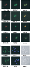
Co-localization of NOP132 with DDX47, DDX18, RPL3, and NOP254 in the nucleolus. (a) BHK21 cells were transiently cotransfected with DsRed-NOP132 (red) and DDX47-EGFP, DDX18-EGFP, RPL3-EGFP, or KIAA0539/NOP254-EGFP (green). The cells were fixed and processed for confocal microscopy imaging as described in the Materials and Methods. Scale bars, 10 µm. (b) HeLa cells were fixed and immunostained with affinity purified rabbit polyclonal anti-DDX18 or anti-DDX47 followed by fluorescein-isothiocyanate-conjugated anti-rabbit secondary antibody. DNA was stained with Hoechst dye. Phase images are shown in the right panels.
Figure 4
Figure 4
Association of DDX47 with pre-rRNAs. (a) HeLa cells were transiently transfected with two dsRNAs for each RNAi experiment as described in the Materials and Methods. WT, Control fruit fly luciferase oligonucleotide; DDX47 RNAi, oligonucleotides DDX47-1 and -2; NOP132 RNAi, oligonucleotides NOP132-B and -C. After transfection for 1 d, total RNAs were purified and processed for Northern blot analysis using ITS1, ITS2, 28S, 18S, 5.8S, or α-actin DNAs as probes (shown in parentheses). RNA from control (WT), NOP132 RNAi-treated, and DDX47 RNAi-treated cells was electrophoresed on a formaldehyde agarose gel as described in the Materials and Methods. Radioactivity in each lane was measured using the Fuji BAS2500 Image Analyzer (Fuji Photo Film Co. Ltd, Japan). Relative radioactivity of each rRNA or rRNA precursor is shown at the bottom of the panels. (b) Proteins from the siRNA-treated cells were isolated and processed for western blotting analysis using anti-NOP132N, anti-DDX47, or anti-RAN as a loading control. (c) 293 cells were transiently transfected with FLAG- DDX47. After transfection for 2 d, a cell lysate was prepared and the DDX47-associating complex was purified with anti-FLAG. RNA was purified from the immunoprecipitate and processed for Northern blot analysis using ITS1, ITS2, 28S, 5.8S, or 18S oligonucleotide probes. As a control, an untransfected 293 cell lysate was prepared and processed as above. Relative radioactivity of each rRNA or rRNA precursor is shown at the bottom of the panels. (d) Schematic representation of the pre-rRNAs detected in the Northern blots. Possible points where DDX47 might act are indicated in the figure. The pre-rRNA cleavage sites (1- 3, 3′, 4′) are indicated by arrowheads.
Figure 5
Figure 5
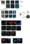
NOP132 is required for DDX47 to properly localize to the nucleolus. Localization of DDX47 by anti-DDX47 immunofluorescence in HeLa cells treated with NOP132 siRNAs to reduce expression of NOP132. Control cells were transfected with luciferase siRNA duplex. (a) NOP132 RNAi in HeLa cells (upper panels). Control luciferase RNAi in HeLa cells (lower panels). Confocal images were taken with the Olympus laser-scanning confocal microscope LSM-GB200 system (left panel). DNA was stained with DAPI. Merge images of DDX47 and DNA are shown in the rightmost panels. Fluorescence images were taken with the Zeiss Axiophot microscope (right panels). Scale bars, 10 µm. (b) HeLa cells were treated without (control, lower panels) or with 5 µg/ml actinomycin D for 8 h (upper panels) and then immunostained with anti-DDX47 and stained with DAPI. Left panels, confocal fluorescence microscopy. Right panels, fluorescence microscopy. (c) A schematic model of the change in the localization of DDX47. In the untreated state, DDX47 (in black) is localized to the nucleolus. (d) 293 cells were transiently cotransfected with vectors carrying EGFP-DDX47 and FLAG-NOP132 coiled-coil domain minus a NLS (residues 667–960; FLAG-Coil). The cells were fixed and stained with anti-FLAG followed by Alexa Fluor 594-goat anti-mouse IgG. Upper and middle panels, fluorescence microscopy; lower panels, confocal fluorescence microscopy. Arrowheads indicate cells in which the EGFP-DDX47 protein distributed evenly in the nucleus. DNA was stained with Hoechst dye. (e) 293 cells were transiently transfected with the NOP132 FLAG-Coil vector. The cells were fixed and stained with FLAG monoclonal antibody and then with Alexa Fluor 594-goat anti-mouse IgG. DNA was stained with Hoechst dye. Fluorescence images were taken with the Zeiss Axiophot microscope. (f) 293 cells were transiently transfected with EGFP-DDX47 vector. DNA was stained with Hoechst dye. Fluorescence images were taken with the Zeiss Axiophot microscope.
Figure 6
Figure 6
Analysis of regions responsible for subcellular localization of DDX47. (a) Serial deletion constructs of EGFP-fused DDX47 were used to transfect BHK21 cells. Subcellular localization of all constructs was determined by fluorescence microscopy. Representative fluorescence images are shown. Interaction of DDX47 deletion constructs with NOP132 was determined using two different yeast two-hybrid read-outs, a chromogenic β-galactosidase assay and a colony growth assay, and the results are shown at the far right. Domains: DEADc, CDD accession no. cd00268 (38); HELICc, CDD accession no. cd00079. Abbreviations: N, nucleus/nucleolus; C, cytoplasm; NES, nuclear export signal; NLS, nuclear localization signal; +, yeast two-hybrid interaction between the DDX47 deletion construct and NOP132; −, no interaction. Hatched box indicates the NOP132-interacting region. N, nucleus; C, cytoplasm. (b) DDX47 amino acid sequence is shown indicating possible NES and NLS regions (underlined) and motifs (bold). (c) Sequence alignment of the DDX47 and HELICc motifs. Conserved RNA helicase DEAD-box motifs IV, V, and VI are indicated (48). Two dots indicate identity and a single dot indicates chemically similar amino acids.
Figure 7
Figure 7
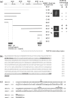
Determination of NOP132-interacting region of DDX47. (a) Summary of the region of NOP132 that associates with DDX47 and DDX18. The three coiled-coil structures are shown as black bars. (b) Deletion constructs of NOP132 in the vector pACT2 were examined for their interaction with DDX47 or DDX18 in the vector pAS1 using a yeast two-hybrid assay. Right panels show the results of the β-galactosidase filter assay and colony growth assay. β-gal activity indicates that an interaction had occurred. Abbreviations: + yeast two-hybrid interaction between NOP132 deletion construct and DDX47 or DDX18; +/− weak interaction; − no interaction. (c) Sequence alignment of the C terminus of DDX18 and DDX47 showing 37.3% identity between these proteins. Two dots indicate identity and a single dot indicates chemically similar amino acids.

Similar articles

Cited by

References

    1. Granneman S., Baserga S.J. Ribosome biogenesis: of knobs and RNA processing. Exp. Cell Res. 2004;296:43–50. - PubMed
    1. Fromont-Racine M., Senger B., Saveanu C., Fasiolo F. Ribosome assembly in eukaryotes. Gene. 2003;313:17–42. - PubMed
    1. Venema J., Tollervey D. Ribosome synthesis in Saccharomyces cerevisiae. Annu. Rev. Genet. 1999;33:261–311. - PubMed
    1. Takahashi N., Yanagida M., Fujiyama S., Hayano T., Isobe T. Proteomic snapshot analyses of preribosomal ribonucleoprotein complexes formed at various stages of ribosome biogenesis in yeast and mammalian cells. Mass Spectrom Rev. 2003;22:287–317. - PubMed
    1. Nishimoto T. Upstream and downstream of ran GTPase. Biol. Chem. 2000;381:397–405. - PubMed

Publication types

MeSH terms