. 2006;34(16):4622-9.
doi: 10.1093/nar/gkl458. Epub 2006 Sep 8.

Characterization of DGCR8/Pasha, the essential cofactor for Drosha in primary miRNA processing

Affiliations

Characterization of DGCR8/Pasha, the essential cofactor for Drosha in primary miRNA processing

Kyu-Hyeon Yeom et al. Nucleic Acids Res. 2006.

Abstract

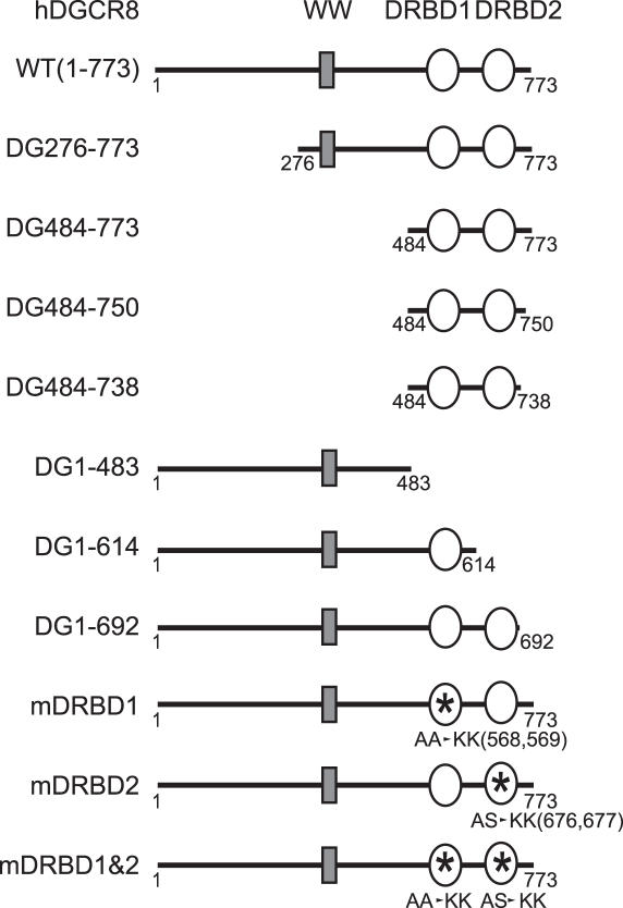
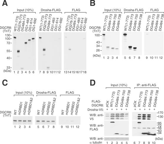
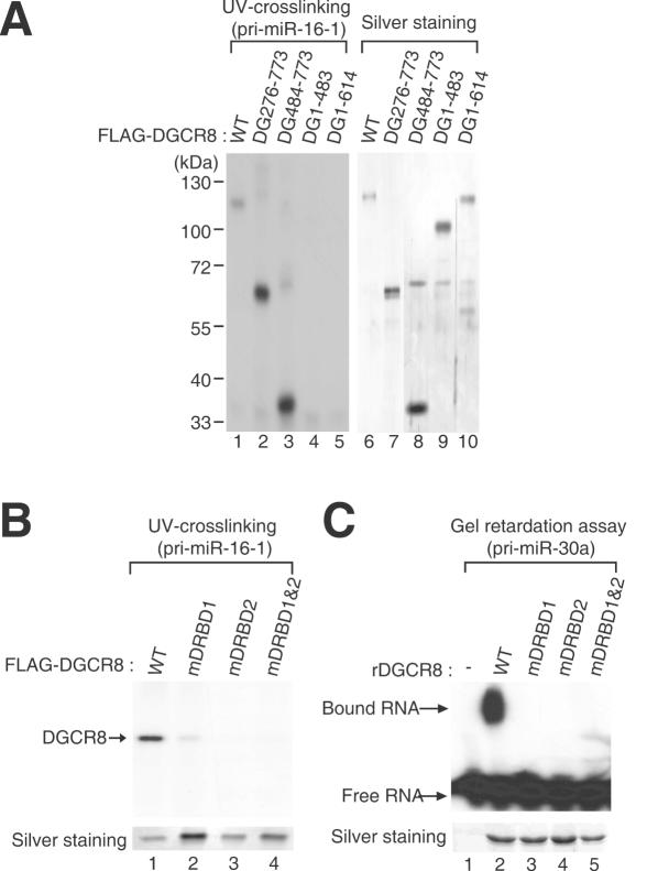
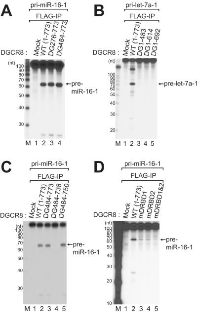
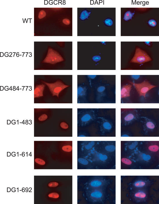
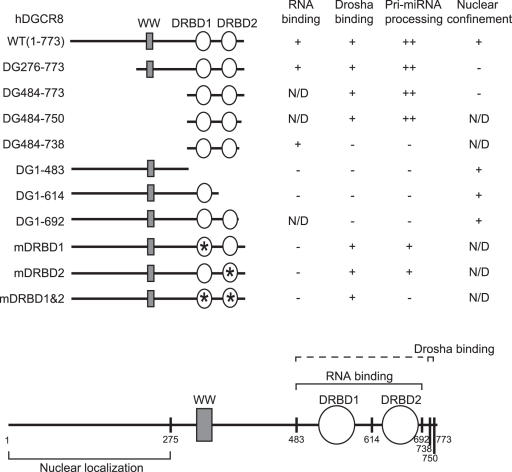
DGCR8/Pasha is an essential cofactor for Drosha, a nuclear RNase III that cleaves the local hairpin structures embedded in long primary microRNA transcripts (pri-miRNAs) in eukaryotes. Although our knowledge of pri-miRNA processing has significantly advanced in recent years, the precise role of DGCR8 in this pathway remains unclear. In our present study, we dissect the domains in DGCR8 that contribute to the processing of pri-miRNAs and the subcellular localization of DGCR8. Drosha is stabilized through an interaction between its middle domain and the conserved C-terminal domain of DGCR8. Furthermore, DGCR8, but not Drosha, can directly and stably interact with pri-miRNAs, and the tandem dsRNA-binding domains (dsRBDs) in DGCR8 are responsible for this recognition. Moreover, the DGCR8 N-terminal region upstream of its dsRBDs is unnecessary for pri-miRNA processing but is critical for nuclear localization. Our study thus provides further insights into the mechanism of action of the Drosha-DGCR8 complex in pri-miRNA processing.


Figures

Figure 1
Figure 1
DGCR8 mutants used in this study. Asterisks represent the sites of point mutagenesis.
Figure 2
Figure 2
Identification of the Drosha-interacting domain. (A–C) In vitro binding assay. DGCR8 wild-type and the indicated mutant proteins were synthesized by TnT (in vitro transcription and translation, Invitrogen) reactions and incubated with the Drosha-FLAG protein immobilized on anti-FLAG antibody that is conjugated on agarose beads. The left panels show 10% of the input proteins used in the binding reactions, whereas the middle panels visualize the proteins interacting with Drosha-FLAG. As a control (right panels, pull-down with FLAG), an empty vector expressing FLAG tag only was transfected and the cell lysates were used for immunoprecipitation. (D) Immunoprecipitation followed by western blot analysis. FLAG-DGCR8 mutant constructs were co-expressed with Drosha-V5. Immunoprecipitation was performed with anti-FLAG antibody and western blotting was carried out using either anti-V5 antibody (upper panels) or anti-FLAG antibody (lower panels).
Figure 3
Figure 3
DGCR8 domains responsible for pri-miRNA binding. (A) UV-crosslinking experiments between a series of DGCR8 deletion mutants and 32P-UTP radiolabeled pri-miR-16-1. After UV-crosslinking, residual RNA was removed by treatment with an RNase A/T1 mix. The left panel visualizes the radioactivity associated with recombinant FLAG-DGCR8 proteins, whereas the right panel indicates the amounts of protein used in this experiment as visualized by silver staining. (B) UV-crosslinking between a series of DGCR8 point mutants and 32P-UTP radiolabeled pri-miR-16-1. The upper panel displays the radioactivity crosslinked to the immunopurified FLAG-DGCR8 proteins and the lower panel indicates the amount of protein used in this experiment as determined by silver staining. (C) Gel retardation assay of pri-miR-30a binding to DGCR8 point mutants. Recombinant DGCR8 proteins prepared from E.coli were pre-treated with thrombin to separate the GST tag prior to the assay. The lower panel shows silver stained gel of recombinant DGCR8 protein used in this experiment.
Figure 4
Figure 4
DGCR8 domains required for pri-miRNA processing. In vitro pri-miRNA processing assay of DGCR8 mutants. FLAG-DGCR8 was immunoprecipitated using anti-FLAG antibody-conjugated agarose beads in low salt buffer (buffer D-K′200), and subsequently subjected to an in vitro pri-miRNA processing assay. Pri-miR-16-1 or pri-let-7a-1, labeled internally with 32P-UTP, was used as the substrate. Decade markers (Ambion) were used as the size markers.
Figure 5
Figure 5
Subcellular localization of DGCR8 and its deletion mutants. HeLa cells were transiently transfected with V5-tagged constructs, including WT DGCR8, and the DG276–773, DG484–773, DG1–483, DG1–614 and DG1–692 mutants. The left panels show DGCR8 protein signals visualized with rabbit anti-V5 serum (1:500) and Alexa Flour 594 goat anti-rabbit IgG (1:400). The middle panels show DAPI staining of the nucleus and the right panels display an overlay of these two signals.
Figure 6
Figure 6
Summary of the DGCR8 mutagenesis findings. Our DGCR8 mutants can be characterized into four different groups; pri-miRNA binding, Drosha-binding, pri-miRNA processing and nuclear confinement activity. N/D, not determined.

Similar articles

Cited by

References

    1. Bartel D.P. MicroRNAs: genomics, biogenesis, mechanism, and function. Cell. 2004;116:281–297. - PubMed
    1. Kim V.N. Small RNAs: Classification, Biogenesis, and Function. Mol. Cells. 2005;19:1–15. - PubMed
    1. Xie Z., Allen E., Fahlgren N., Calamar A., Givan S.A., Carrington J.C. Expression of Arabidopsis MIRNA genes. Plant Physiol. 2005;138:2145–2154. - PMC - PubMed
    1. Lewis B.P., Burge C.B., Bartel D.P. Conserved seed pairing, often flanked by adenosines, indicates that thousands of human genes are microRNA targets. Cell. 2005;120:15–20. - PubMed
    1. Croce C.M., Calin G.A. miRNAs, cancer, and stem cell division. Cell. 2005;122:6–7. - PubMed

Publication types