

Disease-causing mutations in proteins: structural analysis of the CYP1B1 mutations causing primary congenital glaucoma in humans
- PMID: 16963504
- PMCID: PMC1779944
- DOI: 10.1529/biophysj.106.085498
Disease-causing mutations in proteins: structural analysis of the CYP1B1 mutations causing primary congenital glaucoma in humans
Abstract
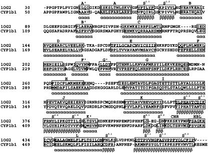
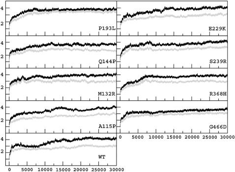
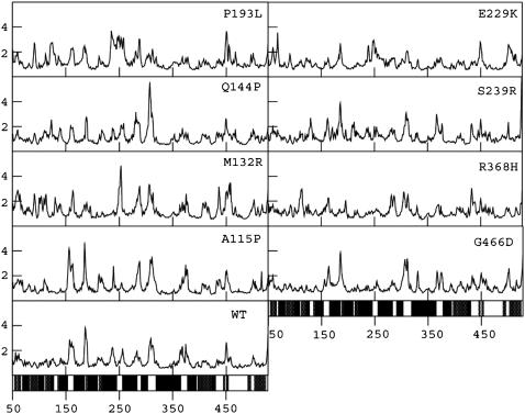
In this communication, we report an in-depth structure-based analysis of the human CYP1b1 protein carrying disease-causing mutations that are discovered in patients suffering from primary congenital glaucoma (PCG). The "wild-type" and the PCG mutant structures of the human CYP1b1 protein obtained from comparative modeling were subjected to long molecular dynamics simulations with an intention of studying the possible impact of these mutations on the protein structure and hence its function. Analysis of time evolution as well as time averaged values of various structural properties--especially of those of the functionally important regions: the heme binding region, substrate binding region, and substrate access channel--gave some insights into the possible structural characteristics of the disease mutant and the wild-type forms of the protein. In a nutshell, compared to the wild-type the core regions in the mutant structures are associated with subtle but significant changes, and the functionally important regions seem to adopt such structures that are not conducive for the wild-type-like functionality.
Figures
Similar articles
-
Characterization of the biochemical and structural phenotypes of four CYP1B1 mutations observed in individuals with primary congenital glaucoma.Pharmacogenet Genomics. 2008 Aug;18(8):665-76. doi: 10.1097/FPC.0b013e3282ff5a36. Pharmacogenet Genomics. 2008. PMID: 18622259
-
Effects of disease causing mutations on the essential motions in proteins.J Biomol Struct Dyn. 2009 Apr;26(5):609-24. doi: 10.1080/07391102.2009.10507276. J Biomol Struct Dyn. 2009. PMID: 19236111
-
Novel cytochrome P4501B1 (CYP1B1) gene mutations in Japanese patients with primary congenital glaucoma.Invest Ophthalmol Vis Sci. 2001 Sep;42(10):2211-6. Invest Ophthalmol Vis Sci. 2001. PMID: 11527932
-
Molecular genetics of primary congenital glaucoma.Eye (Lond). 2000 Jun;14 ( Pt 3B):422-8. doi: 10.1038/eye.2000.126. Eye (Lond). 2000. PMID: 11026969 Review.
-
CYP1B1, a developmental gene with a potential role in glaucoma therapy.Xenobiotica. 2009 Aug;39(8):606-15. doi: 10.1080/00498250903000198. Xenobiotica. 2009. PMID: 19622003 Review.
Cited by
-
Zebrafish Cyp1b1 knockout alters eye and brain metabolomic profiles, affecting ocular and neurobehavioral function.Toxicol Appl Pharmacol. 2025 Mar;496:117246. doi: 10.1016/j.taap.2025.117246. Epub 2025 Jan 29. Toxicol Appl Pharmacol. 2025. PMID: 39890032 Free PMC article.
-
Novel CYP1B1 mutations in consanguineous Pakistani families with primary congenital glaucoma.Mol Vis. 2008;14:2002-9. Epub 2008 Nov 3. Mol Vis. 2008. PMID: 18989382 Free PMC article.
-
A novel 9-bp insertion detected in steroid 21-hydroxylase gene (CYP21A2): prediction of its structural and functional implications by computational methods.J Biomed Sci. 2009 Jan 8;16(1):3. doi: 10.1186/1423-0127-16-3. J Biomed Sci. 2009. PMID: 19272182 Free PMC article.
-
Biological roles of cytochrome P450 1A1, 1A2, and 1B1 enzymes.Arch Pharm Res. 2021 Jan;44(1):63-83. doi: 10.1007/s12272-021-01306-w. Epub 2021 Jan 23. Arch Pharm Res. 2021. PMID: 33484438 Review.
-
Mutation screening of the CYP1B1 gene reveals thirteen novel disease-causing variants in consanguineous Pakistani families causing primary congenital glaucoma.PLoS One. 2022 Sep 9;17(9):e0274335. doi: 10.1371/journal.pone.0274335. eCollection 2022. PLoS One. 2022. PMID: 36083974 Free PMC article.
References
-
- deLuise, V. P., and D. R. Anderson. 1983. Primary infantile glaucoma (congenital glaucoma). Surv. Ophthalmol. 28:1–19. - PubMed
-
- Gencik, A., A. Gencikova, and V. Ferak. 1982. Population genetical aspects of primary congenital glaucoma. I. Incidence, prevalence, gene frequency, and age of onset. Hum. Genet. 61:193–197. - PubMed
-
- Sarfarazi, M., A. N. Akarsu, A. Hossain, M. E. Turacli, S. G. Aktan, M. Barsoum-Homsy, L. Chevrette, and B. S. Sayli. 1995. Assignment of a locus (GLC3A) for primary congenital glaucoma (Buphthalmos) to 2p21 and evidence for genetic heterogeneity. Genomics. 30:171–177. - PubMed
-
- Akarsu, A. N., M. E. Turacli, S. G. Aktan, M. Barsoum-Homsy, L. Chevrette, B. S. Sayli, and M. Sarfarazi. 1996. A second locus (GLC3B) for primary congenital glaucoma (Buphthalmos) maps to the 1p36 region. Hum. Mol. Genet. 5:1199–1203. - PubMed
-
- Stoilov, I. R. S. M. 2002. The third genetic locus (GLC3C) for primary congenital glaucoma (PCG) maps to Chromosome 14q24.3. Invest. Ophthalmol. Vis. Sci. 43:3015. (Abstr.) - PubMed
Publication types
MeSH terms
Substances
LinkOut - more resources
Full Text Sources
Medical
