

Use of native lactococci as vehicles for delivery of DNA into mammalian epithelial cells
- PMID: 16963550
- PMCID: PMC1636207
- DOI: 10.1128/AEM.01325-06
Use of native lactococci as vehicles for delivery of DNA into mammalian epithelial cells
Abstract
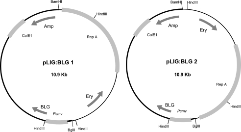
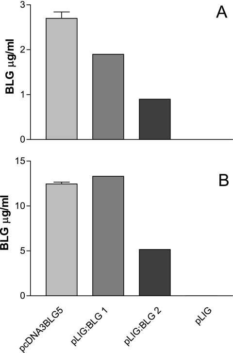
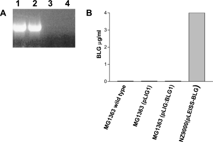
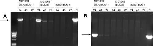
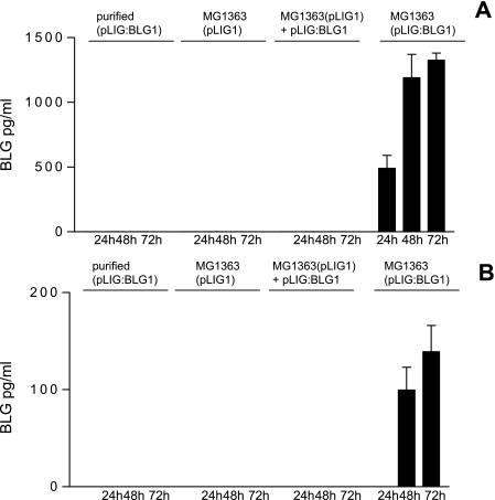
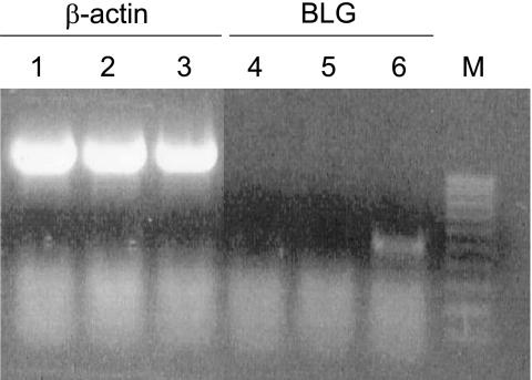
The use of the food-grade bacterium Lactococcus lactis as a DNA delivery vehicle at the mucosal level is an attractive DNA vaccination strategy. Previous experiments showed that recombinant L. lactis expressing the Listeria monocytogenes inlA gene can deliver a functional gene into mammalian cells. Here, we explored the potential use of noninvasive L. lactis strains as a DNA delivery vehicle. We constructed two Escherichia coli-L. lactis shuttle plasmids, pLIG:BLG1 and pLIG:BLG2, containing a eukaryotic expression cassette with the cDNA of bovine beta-lactoglobulin (BLG). The greatest BLG expression after transfection of Cos-7 cells was obtained with pLIG:BLG1, which was then used to transform L. lactis MG1363. The resulting L. lactis strain MG1363(pLIG:BLG1) was not able to express BLG. The potential of L. lactis as a DNA delivery vehicle was analyzed by detection of BLG in Caco-2 human colon carcinoma cells after 3 h of coincubation with (i) purified pLIG:BLG1, (ii) MG1363(pLIG:BLG1), (iii) a mix of MG1363(pLIG) and purified pLIG:BLG1, and (iv) MG1363. Both BLG cDNA and BLG expression were detected only in Caco-2 cells coincubated with MG1363(pLIG:BLG1). There was a decrease in the BLG cDNA level in Caco-2 cells between 24 and 48 h after coincubation. BLG expression by Caco-2 cells started at 24 h and increased between 24 and 72 h. BLG secretion by Caco-2 cells started 48 h after coincubation with MG1363(pLIG:BLG1). We conclude that lactococci can deliver BLG cDNA into mammalian epithelial cells, demonstrating their potential to deliver in vivo a DNA vaccine.
Figures
Similar articles
-
In vitro and in vivo characterization of DNA delivery using recombinant Lactococcus lactis expressing a mutated form of L. monocytogenes Internalin A.BMC Microbiol. 2012 Dec 19;12:299. doi: 10.1186/1471-2180-12-299. BMC Microbiol. 2012. PMID: 23253484 Free PMC article.
-
In vivo transfer of plasmid from food-grade transiting lactococci to murine epithelial cells.Gene Ther. 2008 Aug;15(16):1184-90. doi: 10.1038/gt.2008.59. Epub 2008 Apr 17. Gene Ther. 2008. PMID: 18418419
-
Internalin-expressing Lactococcus lactis is able to invade small intestine of guinea pigs and deliver DNA into mammalian epithelial cells.Microbes Infect. 2005 May;7(5-6):836-44. doi: 10.1016/j.micinf.2005.02.012. Epub 2005 Apr 14. Microbes Infect. 2005. PMID: 15878681
-
Lactococci and lactobacilli as mucosal delivery vectors for therapeutic proteins and DNA vaccines.Microb Cell Fact. 2011 Aug 30;10 Suppl 1(Suppl 1):S4. doi: 10.1186/1475-2859-10-S1-S4. Epub 2011 Aug 30. Microb Cell Fact. 2011. PMID: 21995317 Free PMC article. Review.
-
Engineering lactococci and lactobacilli for human health.Curr Opin Microbiol. 2013 Jun;16(3):278-83. doi: 10.1016/j.mib.2013.06.002. Epub 2013 Jul 11. Curr Opin Microbiol. 2013. PMID: 23850097 Review.
Cited by
-
Lactococcus lactis engineered to deliver hCAP18 cDNA alleviates DNBS-induced colitis in C57BL/6 mice by promoting IL17A and IL10 cytokine expression.Sci Rep. 2022 Sep 19;12(1):15641. doi: 10.1038/s41598-022-19455-3. Sci Rep. 2022. PMID: 36123355 Free PMC article.
-
Recombinant Invasive Lactococcus lactis Carrying a DNA Vaccine Coding the Ag85A Antigen Increases INF-γ, IL-6, and TNF-α Cytokines after Intranasal Immunization.Front Microbiol. 2017 Jul 11;8:1263. doi: 10.3389/fmicb.2017.01263. eCollection 2017. Front Microbiol. 2017. PMID: 28744263 Free PMC article.
-
A novel plasmid for delivering genes into mammalian cells with noninvasive food and commensal lactic acid bacteria.Plasmid. 2011 Jan;65(1):8-14. doi: 10.1016/j.plasmid.2010.09.001. Epub 2010 Sep 9. Plasmid. 2011. PMID: 20832422 Free PMC article.
-
Live Bacterial Vectors-A Promising DNA Vaccine Delivery System.Med Sci (Basel). 2018 Mar 23;6(2):27. doi: 10.3390/medsci6020027. Med Sci (Basel). 2018. PMID: 29570602 Free PMC article. Review.
-
Inhibition of Staphylococcus aureus invasion into bovine mammary epithelial cells by contact with live Lactobacillus casei.Appl Environ Microbiol. 2013 Feb;79(3):877-85. doi: 10.1128/AEM.03323-12. Epub 2012 Nov 26. Appl Environ Microbiol. 2013. PMID: 23183972 Free PMC article.
References
-
- Adel-Patient, K., C. Creminon, D. Boquet, J. M. Wal, and J. M. Chatel. 2001. Genetic immunisation with bovine beta-lactoglobulin cDNA induces a preventive and persistent inhibition of specific anti-BLG IgE response in mice. Int. Arch. Allergy Immunol. 126:59-67. - PubMed
-
- Adel-Patient, K., S. Ah-Leung, C. Creminon, S. Nouaille, J. M. Chatel, P. Langella, and J. M. Wal. 2005. Oral administration of recombinant Lactococcus lactis expressing bovine beta-lactoglobulin partially prevents mice from sensitization. Clin. Exp. Allergy 35:539-546. - PubMed
-
- Avall-Jaaskelainen, S., A. Lindholm, and A. Palva. 2003. Surface display of the receptor-binding region of the Lactobacillus brevis S-layer protein in Lactococcus lactis provides nonadhesive lactococci with the ability to adhere to intestinal epithelial cells. Appl. Environ. Microbiol. 69:2230-2236. - PMC - PubMed
Publication types
MeSH terms
Substances
LinkOut - more resources
Full Text Sources
Other Literature Sources
