AL-57, a ligand-mimetic antibody to integrin LFA-1, reveals chemokine-induced affinity up-regulation in lymphocytes
- PMID: 16963559
- PMCID: PMC1599901
- DOI: 10.1073/pnas.0605716103
AL-57, a ligand-mimetic antibody to integrin LFA-1, reveals chemokine-induced affinity up-regulation in lymphocytes
Abstract
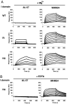
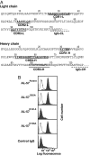
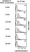
Affinity of integrin lymphocyte function-associated antigen 1 (LFA-1) is enhanced by conformational changes from the low-affinity closed form to the high-affinity (HA) open form of the ligand-binding inserted (I) domain as shown by work with purified I domains. However, affinity up-regulation of LFA-1 on the cell surface by physiological agonists such as chemokines has yet to be demonstrated by monovalent reagents. We characterize a mAb, AL-57 (activated LFA-1 clone 57), that has been developed by phage display that selectively targets the HA open conformation of the LFA-1 I domain. AL-57 discriminates among low-affinity, intermediate-affinity, and HA states of LFA-1. Furthermore, AL-57 functions as a ligand mimetic that binds only upon activation and requires Mg2+ for binding. Compared with the natural ligand intercellular adhesion molecule-1, AL-57 shows a tighter binding to the open I domain and a 250-fold slower off rate. Monovalent Fab AL-57 demonstrates affinity increases on a subset (approximately 10%) of lymphocyte cell surface LFA-1 molecules upon stimulation with CXCL-12 (CXC chemokine ligand 12). Affinity up-regulation correlates with global conformational changes of LFA-1 to the extended form. Affinity increase stimulated by CXCL-12 is transient and peaks 2 to 5 min after stimulation.
Conflict of interest statement
Conflict of interest statement: No conflicts declared.
Figures


Similar articles
-
Effects of I domain deletion on the function of the beta2 integrin lymphocyte function-associated antigen-1.Mol Biol Cell. 2000 Feb;11(2):677-90. doi: 10.1091/mbc.11.2.677. Mol Biol Cell. 2000. PMID: 10679023 Free PMC article.
-
The I domain of integrin leukocyte function-associated antigen-1 is involved in a conformational change leading to high affinity binding to ligand intercellular adhesion molecule 1 (ICAM-1).J Biol Chem. 1998 Oct 16;273(42):27396-403. doi: 10.1074/jbc.273.42.27396. J Biol Chem. 1998. PMID: 9765268
-
2E8 binds to the high affinity I-domain in a metal ion-dependent manner: a second generation monoclonal antibody selectively targeting activated LFA-1.J Biol Chem. 2010 Oct 22;285(43):32860-32868. doi: 10.1074/jbc.M110.111591. Epub 2010 Aug 19. J Biol Chem. 2010. PMID: 20724473 Free PMC article.
-
[An update on beta2 integrin LFA-1 and ligand ICAM-1 signaling].Zhongguo Shi Yan Xue Ye Xue Za Zhi. 2008 Feb;16(1):213-6. Zhongguo Shi Yan Xue Ye Xue Za Zhi. 2008. PMID: 18315934 Review. Chinese.
-
Integrin inside-out signaling and the immunological synapse.Curr Opin Cell Biol. 2012 Feb;24(1):107-15. doi: 10.1016/j.ceb.2011.10.004. Epub 2011 Nov 28. Curr Opin Cell Biol. 2012. PMID: 22129583 Free PMC article. Review.
Cited by
-
Identification, characterization, and epitope mapping of human monoclonal antibody J19 that specifically recognizes activated integrin α4β7.J Biol Chem. 2012 May 4;287(19):15749-59. doi: 10.1074/jbc.M112.341263. Epub 2012 Mar 14. J Biol Chem. 2012. PMID: 22418441 Free PMC article.
-
Structural basis of integrin regulation and signaling.Annu Rev Immunol. 2007;25:619-47. doi: 10.1146/annurev.immunol.25.022106.141618. Annu Rev Immunol. 2007. PMID: 17201681 Free PMC article. Review.
-
Structural surfaceomics reveals an AML-specific conformation of integrin β2 as a CAR T cellular therapy target.Nat Cancer. 2023 Nov;4(11):1592-1609. doi: 10.1038/s43018-023-00652-6. Epub 2023 Oct 30. Nat Cancer. 2023. PMID: 37904046 Free PMC article.
-
Identification and X-ray co-crystal structure of a small-molecule activator of LFA-1-ICAM-1 binding.Angew Chem Int Ed Engl. 2014 Apr 22;53(17):4322-6. doi: 10.1002/anie.201310240. Epub 2014 Apr 1. Angew Chem Int Ed Engl. 2014. PMID: 24692345 Free PMC article.
-
Selective gene silencing in activated leukocytes by targeting siRNAs to the integrin lymphocyte function-associated antigen-1.Proc Natl Acad Sci U S A. 2007 Mar 6;104(10):4095-100. doi: 10.1073/pnas.0608491104. Epub 2007 Feb 28. Proc Natl Acad Sci U S A. 2007. PMID: 17360483 Free PMC article.
References
-
- Springer TA. Cell. 1994;76:301–314. - PubMed
-
- Dustin ML, Springer TA. In: Guidebook to the Extracellular Matrix and Adhesion Proteins. Kreis T, Vale R, editors. New York: Sambrook & Tooze; 1999. pp. 228–232.
Publication types
MeSH terms
Substances
Grants and funding
LinkOut - more resources
Full Text Sources
Other Literature Sources

