C-terminal WxL domain mediates cell wall binding in Enterococcus faecalis and other gram-positive bacteria
- PMID: 16963569
- PMCID: PMC1797349
- DOI: 10.1128/JB.00773-06
C-terminal WxL domain mediates cell wall binding in Enterococcus faecalis and other gram-positive bacteria
Abstract
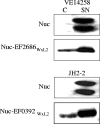
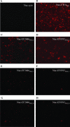
Analysis of the genome sequence of Enterococcus faecalis clinical isolate V583 revealed novel genes encoding surface proteins. Twenty-seven of these proteins, annotated as having unknown functions, possess a putative N-terminal signal peptide and a conserved C-terminal region characterized by a novel conserved domain designated WxL. Proteins having similar characteristics were also detected in other low-G+C-content gram-positive bacteria. We hypothesized that the WxL region might be a determinant of bacterial cell location. This hypothesis was tested by generating protein fusions between the C-terminal regions of two WxL proteins in E. faecalis and a nuclease reporter protein. We demonstrated that the C-terminal regions of both proteins conferred a cell surface localization to the reporter fusions in E. faecalis. This localization was eliminated by introducing specific deletions into the domains. Interestingly, exogenously added protein fusions displayed binding to whole cells of various gram-positive bacteria. We also showed that the peptidoglycan was a binding ligand for WxL domain attachment to the cell surface and that neither proteins nor carbohydrates were necessary for binding. Based on our findings, we propose that the WxL region is a novel cell wall binding domain in E. faecalis and other gram-positive bacteria.
Figures


Similar articles
-
Characterization of Enterococcus faecalis alkaline phosphatase and use in identifying Streptococcus agalactiae secreted proteins.J Bacteriol. 1999 Sep;181(18):5790-9. doi: 10.1128/JB.181.18.5790-5799.1999. J Bacteriol. 1999. PMID: 10482522 Free PMC article.
-
Structural studies of the Enterococcus faecalis SufU [Fe-S] cluster protein.BMC Biochem. 2009 Feb 2;10:3. doi: 10.1186/1471-2091-10-3. BMC Biochem. 2009. PMID: 19187533 Free PMC article.
-
Surface protein EF3314 contributes to virulence properties of Enterococcus faecalis.Int J Artif Organs. 2009 Sep;32(9):611-20. doi: 10.1177/039139880903200910. Int J Artif Organs. 2009. PMID: 19856273 Free PMC article.
-
Cell-cell communication in gram-positive bacteria.Annu Rev Microbiol. 1997;51:527-64. doi: 10.1146/annurev.micro.51.1.527. Annu Rev Microbiol. 1997. PMID: 9343359 Review.
-
[Autolysis of bacterial cell surface and its physiology (biosynthesis)].Tanpakushitsu Kakusan Koso. 1972 Aug;17(8):581-7. Tanpakushitsu Kakusan Koso. 1972. PMID: 4143920 Review. Japanese. No abstract available.
Cited by
-
Overexpression of Enterococcus faecalis elr operon protects from phagocytosis.BMC Microbiol. 2015 May 25;15:112. doi: 10.1186/s12866-015-0448-y. BMC Microbiol. 2015. PMID: 26003173 Free PMC article.
-
Fitness Restoration of a Genetically Tractable Enterococcus faecalis V583 Derivative To Study Decoration-Related Phenotypes of the Enterococcal Polysaccharide Antigen.mSphere. 2019 Jul 10;4(4):e00310-19. doi: 10.1128/mSphere.00310-19. mSphere. 2019. PMID: 31292230 Free PMC article.
-
Microdiversity of Enterococcus faecalis isolates in cases of infective endocarditis: selection of non-synonymous mutations and large deletions is associated with phenotypic modifications.Emerg Microbes Infect. 2021 Dec;10(1):929-938. doi: 10.1080/22221751.2021.1924865. Emerg Microbes Infect. 2021. PMID: 33913790 Free PMC article.
-
Adhesion of the genome-sequenced Lactococcus lactis subsp. cremoris IBB477 strain is mediated by specific molecular determinants.Appl Microbiol Biotechnol. 2016 Nov;100(22):9605-9617. doi: 10.1007/s00253-016-7813-0. Epub 2016 Sep 29. Appl Microbiol Biotechnol. 2016. PMID: 27687992 Free PMC article.
-
Listeria monocytogenes surface proteins: from genome predictions to function.Microbiol Mol Biol Rev. 2007 Jun;71(2):377-97. doi: 10.1128/MMBR.00039-06. Microbiol Mol Biol Rev. 2007. PMID: 17554049 Free PMC article. Review.
References
-
- Bergmann, S., and S. Hammerschmidt. 2006. Versatility of pneumococcal surface proteins. Microbiology 152:295-303. - PubMed
-
- Braun, L., S. Dramsi, P. Dehoux, H. Bierne, G. Lindahl, and P. Cossart. 1997. InlB: an invasion protein of Listeria monocytogenes with a novel type of surface association. Mol. Microbiol. 25:285-294. - PubMed
-
- Burkholder, P. R., and N. H. J. Giles. 1947. Induced biochemical mutations in Bacillus subtilis. Am. J. Bot. 34:345-348. - PubMed
-
- Cabanes, D., P. Dehoux, O. Dussurget, L. Frangeul, and P. Cossart. 2002. Surface proteins and the pathogenic potential of Listeria monocytogenes. Trends Microbiol. 10:238-245. - PubMed
-
- Chaillou, S., M. C. Champomier-Verges, M. Cornet, A. M. Crutz-Le Coq, A. M. Dudez, V. Martin, S. Beaufils, E. Darbon-Rongere, R. Bossy, V. Loux, and M. Zagorec. 2005. The complete genome sequence of the meat-borne lactic acid bacterium Lactobacillus sakei 23K. Nat. Biotechnol. 23:1527-1533. - PubMed
Publication types
MeSH terms
Substances
Associated data
- Actions
LinkOut - more resources
Full Text Sources
Other Literature Sources
Molecular Biology Databases

