. 2006 Nov;188(22):7992-6.
doi: 10.1128/JB.00959-06. Epub 2006 Sep 8.

Cooperative and critical roles for both G domains in the GTPase activity and cellular function of ribosome-associated Escherichia coli EngA

Affiliations

Cooperative and critical roles for both G domains in the GTPase activity and cellular function of ribosome-associated Escherichia coli EngA

Amrita Bharat et al. J Bacteriol. 2006 Nov.

Abstract

To probe the cellular phenotype and biochemical function associated with the G domains of Escherichia coli EngA (YfgK, Der), mutations were created in the phosphate binding loop of each. Neither an S16A nor an S217A variant of G domain 1 or 2, respectively, was able to support growth of an engA conditional null. Polysome profiles of EngA-depleted cells were significantly altered, and His(6)-EngA was found to cofractionate with the 50S ribosomal subunit. The variants were unable to complement the abnormal polysome profile and were furthermore significantly impacted with respect to in vitro GTPase activity. Together, these observations suggest that the G domains have a cooperative function in ribosome stability and/or biogenesis.


Figures
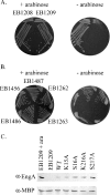
FIG. 1.
FIG. 1.
Complementation of the lethal phenotype of EB1209 by G-domain mutants. (A) The engA diploid strain (EB1208) and the arabinose-inducible null (EB1209) grown on LB-agar-kanamycin in the presence (left) or absence (right) of arabinose. (B) Strains were grown on LB-agar-ampicillin in the presence (left) or absence (right) of 2% (wt/vol) l-Ara. Shown on each plate is EB1209 harboring pDEST14 carrying wild-type engA (EB1487), K15A (EB1262), S16A (EB1263), K216A (EB1486), or S217A (EB1456). (C) Analysis of the expression levels of EngA and its variants from pDEST14 by Western blotting. Lysates obtained from cultures that were grown to an optical density at 600 nm of 0.8 were subjected to Western blot analysis using α-EngA (Cocalico Biologicals, Reamstown, PA) or α-maltose binding protein (New England Biolabs, Beverley, MA) rabbit polyclonal primary antibody and donkey α-rabbit horseradish peroxidase-conjugated secondary antibody. The first and second lanes contain EB1209 grown in the presence or absence of 1% l-Ara, respectively. The remaining lanes contain EB1209 harboring pDEST14 carrying wild-type engA (lane 3), K15A (lane 4), S16A (lane 5), K216A (lane 6), or S217A (lane 7).
FIG. 2.
FIG. 2.
Ribosome profiles of EB1209 with or without complementation. Cells were subjected to one cycle of depletion by growth in the absence of arabinose before growing to an optical density at 600 nm of ∼0.20. Ribosomes were obtained from clarified lysates by centrifugation over a 35% sucrose cushion in buffer A [20 mM Tris, 10.5 mM Mg(OAc)2, 300 mM NH4Cl, 0.5 mM EDTA, 3 mM β-mercaptoethanol, 10 μg/ml RNase-free DNase I]. The ribosomal pellet was analyzed by sedimentation velocity on a Beckman Coulter model XL-I analytical ultracentrifuge. The sedimentation time derivative method (24) was employed, using Microcal Optima v. 6.0 analysis software, to find g(S*), the Gaussian distribution of molecules, at each sedimentation coefficient (S*). Note that S* is a variation of the Svedberg coefficient (S), which is specified at 20°C in water. The effects of temperature and buffer composition on the sedimentation rate were adjusted with the program Sednterp v. 1.01 (D. B. Philo, J. P. Hayes, and T. M. Laue). The profiles of E. coli MG1655 (A), EB1209-1% l-Ara (B), and EB1209 (C) are shown.
FIG. 3.
FIG. 3.
His6-EngA cofractionates predominantly with the large ribosomal subunit. Midexponential phase E. coli BL21(DE3) bacteria harboring (His6)engA-pET28a were induced with 50 μM IPTG (isopropyl-β-d-thiogalactopyranoside) for 30 min at 22°C. The culture was incubated with 200 μg/ml chloramphenicol (FisherBiotech) for 30 s before harvesting. Cleared lysates (13 units of optical density at 260 nm) containing 100 μg/ml chloramphenicol were layered onto a 10 ml gradient of 7% to 47% sucrose in buffer B (10 mM Tris-Cl [pH 7.5], 10 mM MgCl2, 100 mM NH4Cl). Sedimentation of the ribosomes and fractionation of the gradient were carried out as previously described (27). Shown is the profile of absorbance at 254 nm resulting from fractionation. The positions of the 30S and 50S subunits, the 70S monosomes, and the polysomes are indicated. Below the polyribosome trace is an anti-His immunoblot of trichloroacetic acid-precipitated fractions showing the amount of His6-EngA detected. L, 1/100th of the total sample loaded onto the gradient.

Similar articles

Cited by

References

    1. Altschul, S. F., W. Gish, W. Miller, E. W. Myers, and D. J. Lipman. 1990. Basic local alignment search tool. J. Mol. Biol. 215:403-410. - PubMed
    1. Badurina, D. S., M. Zolli-Juran, and E. D. Brown. 2003. CTP:glycerol 3-phosphate cytidylyltransferase (TarD) from Staphylococcus aureus catalyzes the cytidylyl transfer via an ordered Bi-Bi reaction mechanism with micromolar Km values. Biochim. Biophys. Acta 1646:196-206. - PubMed
    1. Bochner, B. R., and B. N. Ames. 1982. Complete analysis of cellular nucleotides by two-dimensional thin layer chromatography. J. Biol. Chem. 257:9759-9769. - PubMed
    1. Bourne, H. R., D. A. Sanders, and F. McCormick. 1991. The GTPase superfamily: conserved structure and molecular mechanism. Nature 349:117-127. - PubMed
    1. Brown, E. D. 2005. Conserved P-loop GTPases of unknown function in bacteria: an emerging and vital ensemble in bacterial physiology. Biochem. Cell Biol. 83:738-746. - PubMed

Publication types

MeSH terms

LinkOut - more resources