. 2006 Nov;188(22):7840-52.
doi: 10.1128/JB.01204-06. Epub 2006 Sep 8.

A conserved tetranucleotide repeat is necessary for wild-type expression of the Moraxella catarrhalis UspA2 protein

Affiliations

A conserved tetranucleotide repeat is necessary for wild-type expression of the Moraxella catarrhalis UspA2 protein

Ahmed S Attia et al. J Bacteriol. 2006 Nov.

Abstract

The UspA2 protein has been shown to be directly involved in the serum-resistant phenotype of Moraxella catarrhalis. The predicted 5'-untranslated regions (UTR) of the uspA2 genes in several different M. catarrhalis strains were shown to contain various numbers (i.e., 6 to 23) of a heteropolymeric tetranucleotide (AGAT) repeat. Deletion of the AGAT repeats from the uspA2 genes in the serum-resistant M. catarrhalis strains O35E and O12E resulted in a drastic reduction in UspA2 protein expression and serum resistance. PCR and transformation were used to construct a series of M. catarrhalis O12E strains that differed only in the number of AGAT repeats in their uspA2 genes. Expression of UspA2 was maximal in the presence of 18 AGAT repeats, although serum resistance attained wild-type levels in the presence of as few as nine AGAT repeats. Increased UspA2 expression was correlated with both increased binding of vitronectin and decreased binding of polymerized C9. Real-time reverse transcription-PCR analysis showed that changes in the number of AGAT repeats affected the levels of uspA2 mRNA, with 15 to 18 AGAT repeats yielding maximal levels. Primer extension analysis indicated that these AGAT repeats were contained in the 5'-UTR of the uspA2 gene. The mRNA transcribed from a uspA2 gene containing 18 AGAT repeats was found to have a longer half-life than that transcribed from a uspA2 gene lacking AGAT repeats. These data confirm that the presence of the AGAT repeats in the 5'-UTR of the uspA2 gene is necessary for both normal expression of the UspA2 protein and serum resistance.


Figures

FIG. 1.
FIG. 1.
PCR and transformation strategies used to construct M. catarrhalis O12E strains with various numbers of AGAT repeats. (A) Chromosomal locus containing the uspA2 gene and flanking DNA. The locations of oligonucleotide primers used for PCRs are shown. (B to D) The different PCR strategies used to construct the strains containing no AGAT repeats (B), 2 AGAT repeats (C), and 6 to 15 AGAT repeats (D) are illustrated.
FIG. 2.
FIG. 2.
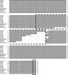
Alignment of the nucleotide sequences of the putative 5′-UTR of the uspA2 genes from 11 different M. catarrhalis strains. Identical nucleotides are shaded in dark gray. This figure was generated by using the CLUSTAL W Alignment program in MacVector (v6.5). The dotted line encloses the AGAT repeats. The small box at the end of these sequences enclosed the ATG translation start codon of the uspA2 gene. The nucleotide sequences of the 5′-UTR from strains O35E, ATCC 25238, and V1171 were derived from a previous study (14).
FIG. 3.
FIG. 3.
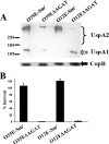
Deletion of the AGAT repeats from the uspA2 gene causes a decrease in both UspA2 protein expression and serum resistance. (A) Western blot analysis of whole-cell lysates of M. catarrhalis strains O35E-Smr, O35EΔAGAT, O12E-Smr, and O12EΔAGAT. The nitrocellulose membrane was probed with MAb 17C7, which recognizes both UspA2 (bracket) and UspA1 (white arrow) (3). The amount of CopB outer membrane protein, as determined by binding of the CopB-reactive MAb 10F3 (21), was used for loading standardization. Protein molecular mass markers (in kilodaltons) are present on the left side of the panel. (B) Serum bactericidal assay with the four strains described in panel A. Bacterial cells were incubated in 10% NHS at 37°C for 30 min. Bacterial aliquots were plated at both t = 0 and t = 30 min. The percent survival was calculated with respect to the original inoculum. These results represent the mean of three independent experiments and the error bars represent the standard deviation.
FIG. 4.
FIG. 4.
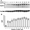
Effect of increasing numbers of AGAT repeats on expression of the UspA2 protein. (A) Western blot analysis of O12E-derived constructs with various numbers of AGAT repeats in their uspA2 genes. Proteins present in whole-cell lysates of these strains were resolved by SDS-PAGE under nonreducing conditions and transferred to nitrocellulose membranes. The membranes were probed with MAb 17C7, which binds the M. catarrhalis UspA1 and UspA2 proteins (3). The region of the gel containing the UspA1 protein is not present in this image. As a loading control, membranes were probed with the CopB-reactive MAb 10F3 (21). (B) Flow cytometric analysis of the reactivity of MAb 17C7 with O12E constructs with various numbers of AGAT repeats in the 5′-UTR of their uspA2 genes. Whole cells of uspA1 mutants of these O12E constructs were probed with MAb 17C7, followed by washing and incubation with a fluorescein isothiocyanate-conjugated antiserum to mouse immunoglobulin G. After a washing step, the cells were analyzed by flow cytometry, and the gmf values were recorded. These results represent the mean of three independent experiments, and the error bars represent the standard deviation.
FIG. 5.
FIG. 5.
Serum bactericidal assay performed with O12E constructs with various numbers of AGAT repeats in their uspA2 genes. Bacterial cells were incubated in 10% NHS (□) and 30% NHS (▪) at 37°C for 30 min. Bacterial aliquots were plated at both t = 0 and t = 30 min. The percent survival was calculated with respect to the original inoculum. These results represent the mean of three independent experiments, and the error bars represent the standard deviation.
FIG. 6.
FIG. 6.
Binding of serum components from NHS to O12E constructs with various numbers of AGAT repeats in their uspA2 genes. Bacterial cells were incubated in 10% NHS at 37°C for 30 min. The cells were then washed and whole-cell lysates were prepared and analyzed by Western blotting. (A) Vitronectin bound to M. catarrhalis cells. Proteins present in the samples were resolved by SDS-PAGE under reducing conditions, transferred to nitrocellulose membranes, and probed with an MAb against human vitronectin. The last lane contains a sample of NHS diluted 1:200. (B) Polymerized C9 bound to M. catarrhalis cells. Proteins present in the samples were resolved by SDS-PAGE under nonreducing conditions, transferred to polyvinylidene difluoride membranes, and probed with an MAb to SC5b-9 that recognizes a neoepitope in polymerized C9 in the membrane attack complex. The last lane of panel A contains a control sample of zymosan-activated NHS (ZAS) that was probed with the same MAb to detect polymerized C9 (5). As a loading control, membranes were probed with M. catarrhalis CopB-reactive MAb 10F3 (21). Protein molecular mass markers (in kilodaltons) are presented on the left side of each panel.
FIG. 7.
FIG. 7.
Real-time RT-PCR analysis of uspA2 gene expression by O12E constructs with various numbers of AGAT repeats in their uspA2 genes. Total RNA isolated from O12E constructs with various numbers of AGAT repeats in their uspA2 genes was used for real-time RT-PCR with primers specific for the uspA2 and copB genes. The data analysis was carried out by using the 7500 System SDS software v.13, applying the relative quantification ΔΔCT method. The level of the uspA2 message was normalized according to the level of the copB message, and the data are presented as a fold increase using the normalized level of the uspA2 gene of O12EΔAGAT as the calibrator. These data represent the mean of three independent experiments (each performed with samples in triplicate), and the error bars represent the standard deviations.
FIG. 8.
FIG. 8.
Determination of the uspA2 transcriptional start point by using primer extension analysis. (A) Primer extension results obtained with RNA isolated from M. catarrhalis O12E-9rpts and AA52-Rev. The arrow indicates the position of the RT product, and the repeat region is indicated with the bracket. The asterisk marks the position from which transcription starts. (B) Nucleotide sequence of the 5′ end of the M. catarrhalis O12E uspA2 gene. The C nucleotide marked with “+1” shows the predicted transcriptional start point determined from the data in panel A. Sequences similar to the −10 and −35 consensus sequences seen in bacterial gene promoters are marked with black bars. The start of the uspA2 ORF is marked with the black arrow, and the AGAT repeat region is contained within the dotted box. The locations of binding sites for the oligonucleotide primers AA52-Rev, AA26-Rev, and AA9-Rev are underlined and labeled as a, b, and c, respectively.
FIG. 9.
FIG. 9.
Stability of the uspA2 and copB transcripts from strains O12EΔAGAT and O12E-18rpts. RNA isolated from M. catarrhalis strains O12E-0rpts and O12E-18rpts at different time points after the addition of rifampin was analyzed by using quantitative real-time RT-PCR to determine the amount of both uspA2 (A) and copB (B) transcripts. The percentage of mRNA left at each time point was determined and plotted. These results are the mean of three experiments, and the error bars indicate the standard deviation. The asterisk indicates that the difference between the two tested strains in the percentage of remaining uspA2 mRNA was statistically significant, with the indicated P value.
FIG. 10.
FIG. 10.
Predicted secondary structure of the RNA transcribed from the 5′-UTR of uspA2 genes with various numbers of AGAT repeats. The Mfold program (36, 56) was used to predict these structures. The arrows indicate the position of the predicted ribosome-binding site in each structure. The ΔG of each secondary structure is listed in kcal/mol.

Similar articles

Cited by

References

    1. Aebi, C., L. D. Cope, J. L. Latimer, S. E. Thomas, C. A. Slaughter, G. H. McCracken, Jr., and E. J. Hansen. 1998. Mapping of a protective epitope of the CopB outer membrane protein of Moraxella catarrhalis. Infect. Immun. 66:540-548. - PMC - PubMed
    1. Aebi, C., E. R. Lafontaine, L. D. Cope, J. L. Latimer, S. R. Lumbley, G. H. McCracken, Jr., and E. J. Hansen. 1998. Phenotypic effect of isogenic uspA1 and uspA2 mutations on Moraxella catarrhalis O35E. Infect. Immun. 66:3113-3119. - PMC - PubMed
    1. Aebi, C., I. Maciver, J. L. Latimer, L. D. Cope, M. K. Stevens, S. E. Thomas, G. H. McCracken, Jr., and E. J. Hansen. 1997. A protective epitope of Moraxella catarrhalis is encoded by two different genes. Infect. Immun. 65:4367-4377. - PMC - PubMed
    1. Attia, A. S., E. R. Lafontaine, J. L. Latimer, C. Aebi, G. A. Syrogiannopoulos, and E. J. Hansen. 2005. The UspA2 protein of Moraxella catarrhalis is directly involved in the expression of serum resistance. Infect. Immun. 73:2400-2410. - PMC - PubMed
    1. Attia, A. S., S. Ram, P. A. Rice, and E. J. Hansen. 2006. Binding of vitronectin by the Moraxella catarrhalis UspA2 protein interferes with late stages of the complement cascade. Infect. Immun. 74:1597-1611. - PMC - PubMed

Publication types

LinkOut - more resources