Review
. 2007 Jan;292(1):H19-27.
doi: 10.1152/ajpheart.00712.2006. Epub 2006 Sep 8.

Preconditioning: a paradigm shift in the biology of myocardial ischemia

Affiliations
Review

Preconditioning: a paradigm shift in the biology of myocardial ischemia

Roberto Bolli. Am J Physiol Heart Circ Physiol. 2007 Jan.

Abstract

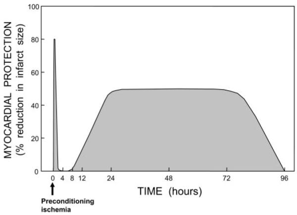
The discovery of preconditioning (PC) has arguably been the single most important development in the field of ischemic biology in the past 20 years. The significance of this phenomenon transcends cardiovascular medicine, since it is ubiquitously observed in virtually every tissue of the body. This article reviews the pathophysiology and molecular basis of myocardial PC, with particular emphasis on the late phase of this cardioprotective adaptation. The article also discusses the exploitation of late PC for the development of novel gene therapy strategies aimed at inducing a permanently preconditioned cardiac phenotype (prophylactic cardioprotection). Besides its conceptual interest, deciphering the mechanism of late PC has considerable therapeutic reverberations, since transfer of the genes that underlie late PC would be expected to emulate the salubrious effects of this response of the heart to stress.


Figures

Fig. 1
Fig. 1
Number of publications dealing with cardiac preconditioning since the initial description of this phenomenon in 1986.
Fig. 2
Fig. 2
Schematic illustration of the 2 phases of ischemic preconditioning (PC). Myocardial protection (e.g., % reduction in infarct size) is represented on the y-axis, and time is represented on the x-axis. After the application of a PC stimulus (in this case, ischemia), there is the rapid development of a preconditioned state that confers powerful protection (up to 80–90% reduction in infarct size in some models). However, the protection is ephemeral, dissipating after 1–2 h. This is followed by the slower development of a late phase of protection, which becomes fully manifested at 24 h after the PC stimulus and persists for ~72 h. Thus the duration of the late phase is 30–50 times longer than that of the early phase of PC.
Fig. 3
Fig. 3
Schematic representation of the cellular mechanisms underlying late PC. A nonlethal cellular stress (e.g., reversible ischemia, heat stress, ventricular pacing, or exercise) causes release of chemical signals [nitric oxide (NO), reactive oxygen species (ROS), adenosine, and possibly opioid receptor agonists] that serve as triggers for the development of late PC. These substances activate a complex signal transduction cascade that includes protein kinase C (PKC; specifically, the ∊-isoform), protein tyrosine kinases (specifically, Src and/or Lck), and probably other as yet unknown kinases. A similar activation of PKC and downstream kinases can be elicited pharmacologically by a wide variety of agents, including naturally occurring—and often noxious—substances (e.g., endotoxin, interleukin-1, TNF-α, TNF-β, leukemia inhibitor factor, or ROS), as well as clinically applicable drugs (NO donors, adenosine A1 or A3 receptor agonists, endotoxin derivatives, or δ1-opioid receptor agonists). The recruitment of PKC and distal kinases leads to activation of NF-κB and almost certainly other transcription factors, resulting in increased transcription of multiple cardioprotective genes and synthesis of multiple cardioprotective proteins that serve as comediators of protection 2–4 days after the PC stimulus. The mediators of late PC identified thus far include inducible nitric oxide synthase (iNOS), cyclooxygenase (COX)-2, heme oxygenase (HO)-1, aldose reductase, extracellular (ec)SOD, and Mn SOD. Among the products of COX-2, PGE2 and/or prostacyclin (PGI2) appear to be the most likely effectors of COX-2-dependent protection. Increased synthesis of heat stress proteins (HSPs) is unlikely to be a mechanism of late PC, although the role of posttranslational modification of preexisting HSPs remains to be determined. In addition, the occurrence of cardioprotection on days 2–4 requires the activity of protein tyrosine kinases and possibly p38 MAPKs, potentially because iNOS and other mediators need to undergo posttranslational modulation to confer protection against ischemia. Opening of ATP-sensitive K+ (KATP) channels is also essential for the protection against infarction (but not against stunning) to become manifest. The exact interrelationships among iNOS, COX-2, HO-1, aldose reductase, ecSOD, Mn SOD, and KATP channels are unclear, although recent evidence suggests that COX-2 is downstream of iNOS (i.e., COX-2 is activated by NO) and NO induces HO-1. eNOS, endothelial NOS.
Fig. 4
Fig. 4
Studies of the role of NO in modulating myocardial ischemia-reperfusion injury. The figure illustrates the studies published in the decade 1990–2000. Of the 92 full-length original manuscripts, 67 concluded that NO is beneficial and only 11 that NO is detrimental (however, several of these “negative” studies have methodological problems). A similar distribution was observed when studies were subdivided according to whether the experiments were performed in vitro (43 studies) or in vivo (49 studies); in both cases, the preponderance of the evidence supports a beneficial role of NO in myocardial ischemia-reperfusion injury. Therefore, the protective effects of NO are not dependent on the use of in vivo or in vitro models. Reprinted from Ref. 3 (Copyright 2001), with permission from Elsevier.
Fig. 5
Fig. 5
Schematic representation of the mechanism of late PC. The PC stimulus activates at least 2 signaling pathways: the PKC∊-Src/Lck-NF-κB pathway and the Janus-activated kinase (JAK)1/JAK2-STAT1/STAT3 pathway. PKC∊ activates the Src and Lck tyrosine kinases, leading to tyrosine phosphorylation of IκBα (the inhibitor of NF-κB); in addition, PKC∊ directly activates IKKα/IKKβ, leading to inhibition of IκBα. NF-κB translocates to the nucleus, where it binds to its cognate sequence on the promoter region of target genes, including iNOS and COX-2. The PC ischemia also activates JAK1 and JAK2, which then tyrosine phosphorylate and activate STAT1 and STAT3. In addition to tyrosine phosphorylation, full activation of STAT1 and STAT3 also requires serine phosphorylation via a PKC∊-Raf-1-MEK1/2-p44/42 MAPK signaling pathway. The activated STAT1/STAT3 heterodimer translocates to the nucleus, where it binds to the promoter of target genes. The combinatorial actions of NF-κB and STAT1/STAT3, and almost certainly other transcription factors, result in transcriptional activation of iNOS, COX-2, HO-1, and ecSOD, leading to the synthesis of the respective proteins. (A multitude of transcription factors are likely to be mobilized during late PC and to lead to the recruitment of cardioprotective genes, acting in concert.) iNOS produces NO, which directly protects the myocardium; in addition, NO activates COX-2, leading to the synthesis of cardioprotective prostanoids, mainly PGI2 and PGE2. It follows from this that the activity of newly synthesized COX-2 protein requires iNOS-dependent NO generation whereas the activity of iNOS does not require COX-2-dependent prostanoid generation; thus COX-2 is downstream of iNOS in the pathophysiological cascade of late PC. In addition, NO can also induce HO-1. Thus iNOS-derived NO can protect the myocardium from recurrent ischemia via direct actions, via activation of COX-2-dependent synthesis of cardioprotective prostanoids, and via generation of HO-1 by-products (CO and biliverdin). Among the products of COX-2, PGE2 and/or PGI2 appear to be the most likely effectors of cytoprotection. A similar upregulation of COX-2 can be elicited pharmacologically by δ-opioid receptor agonists but not by adenosine A1 or A3 receptor agonists.

References

    1. Abete P, Ferrara N, Cioppa A, Ferrara P, Bianco S, Calabrese C, Cacciatore F, Longobardi G, Rengo F. Preconditioning does not prevent postischemic dysfunction in aging heart. J Am Coll Cardiol. 1996;27:1777–1786. - PubMed
    1. Appleby SB, Ristimaki A, Neilson K, Narko K, Hla T. Structure of the human cyclo-oxygenase-2 gene. Biochem J. 1994;302:723–727. - PMC - PubMed
    1. Bolli R. Cardioprotective function of inducible nitric oxide synthase and role of nitric oxide in myocardial ischemia and preconditioning: an overview of a decade of research. J Mol Cell Cardiol. 2001;33:1897–1918. - PubMed
    1. Bolli R. The late phase of preconditioning. Circ Res. 2000;87:972–983. - PubMed
    1. Bolli R, Becker L, Gross G, Mentzer R, Jr, Balshaw D, Lathrop DA. Myocardial protection at a crossroads: the need for translation into clinical therapy. Circ Res. 2004;95:125–134. - PubMed

Publication types

MeSH terms

LinkOut - more resources