. 2007 Feb 22;26(8):1122-36.
doi: 10.1038/sj.onc.1209891. Epub 2006 Sep 11.

The hedgehog regulated oncogenes Gli1 and Gli2 block myoblast differentiation by inhibiting MyoD-mediated transcriptional activation

Affiliations

The hedgehog regulated oncogenes Gli1 and Gli2 block myoblast differentiation by inhibiting MyoD-mediated transcriptional activation

A N Gerber et al. Oncogene. .

Abstract

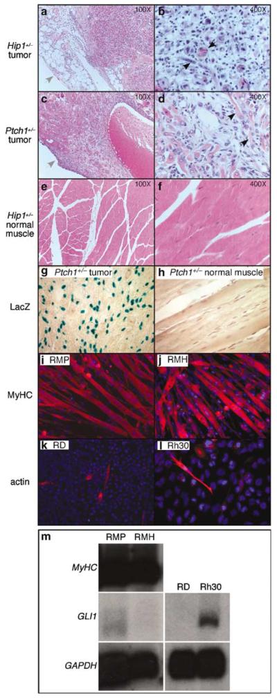
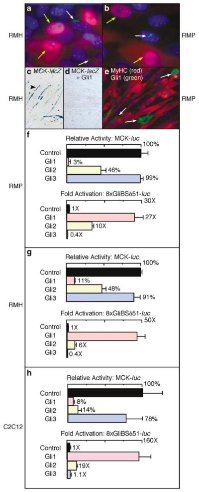
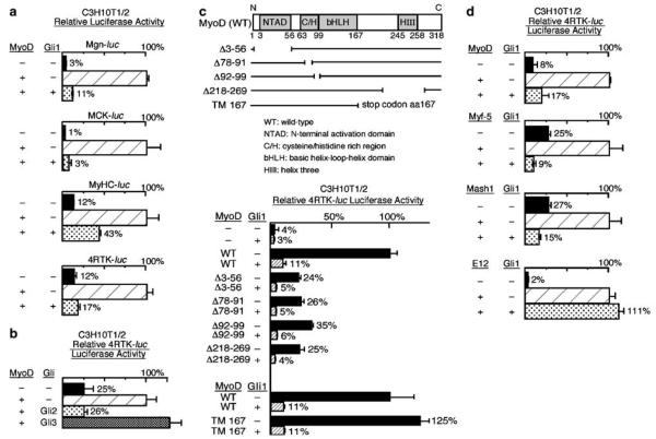
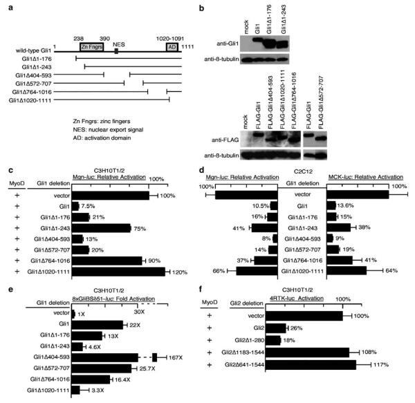
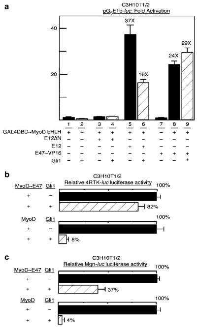
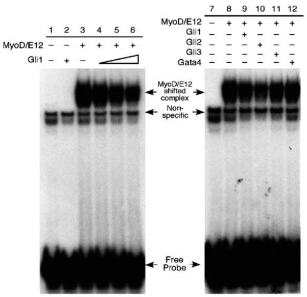
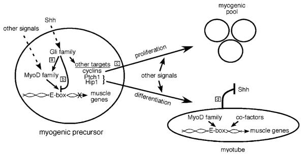
The mechanism by which activation of the Hedgehog (Hh) pathway modulates differentiation and promotes oncogenesis in specific tissues is poorly understood. We therefore, analysed rhabdomyosarcomas from mice that were haploinsufficient for the Hh-binding protein, Hip1, or for the Hh receptor, Patched 1 (Ptch1). Transfection of the Hh-regulated transcription factor Gli1, which is expressed in a subset of mouse and human rhabdomyosarcomas, suppressed differentiation of myogenic rhabdomyosarcoma lines generated from Hip1+/- and Ptch1+/- mice. The closely related factor, Gli2, had similar effects. Gli1 and Gli2 inhibited myogenesis by repressing the capacity of MyoD to activate transcription. Deletion analysis of Gli1 indicated that multiple domains of Gli1 are required for efficient inhibition of MyoD. Gli1 reduced the ability of MyoD to heterodimerize with E12 and bind DNA, providing one mechanism whereby the Gli proteins modulate the activity of MyoD. This novel activity of Gli proteins provides new insights into how Hh signaling modulates terminal differentiation through inhibition of tissue-specific factors such as MyoD. This mechanism may contribute to the broad role of Hh signaling and the Gli proteins in differentiation decisions and cancer formation.


Figures

Figure 1
Figure 1
Analysis of rhabdomyosarcomas from Hip1+/− and Ptch1+/− mice. (a–f) Hematoxylin-and-eosin stained sections of rhabdomyosarcoma isolated from Hip1+/− (a, b) and Ptch1+/− (c, d) mice and of normal muscle from Hip1+/− mice (e, f). (b, d, f) represent higher magnification of (a, c, e). Note the presence of the tumor capsule (indicated by grey arrows in a and c) and the disorganized, highly cellular growth pattern in both tumors (a–d)in comparison to normal muscle (e, f). Histologic features of aberrant skeletal muscle formation, the cardinal feature of rhabdomyosarcoma, were present. For example, black arrows depict multi-nucleated cells (b) and elongated, eosin-positive cells (d). (g, h) X-gal-stained sections of rhabdomyosarcoma and normal muscle isolated from Ptch1+/− mice. In this allele, the β-galactosidase gene was under the control of the Ptch1 promoter (Goodrich et al., 1997) and positive X-gal staining indicates Ptch1 expression. The tumor contained many positively staining cells (blue) not seen in normal muscle. (i–l) Immunohistochemical analysis of myogenic differentiation of rhabdomyosarcoma cell lines (RMH and RMP), derived from Hip1+/− and Ptch1+/− mice, respectively, in comparison to two human rhabdomyosarcoma cell lines, RD and Rh30. The RMP (i) and RMH (j) lines both exhibited extensive myotube formation and expressed myosin heavy chain (MyHC) (red) and sarcomeric actin (not shown), markers for terminal differentiation. In contrast, in the RD (k) and Rh30 (l) lines, only occasional mononucleated cells that expressed sarcomeric actin (red) and MyHC (not shown) were observed. (m) Northern blot analysis of gene expression in the rhabdomyosarcoma cell lines used in this study. The RMH and RMP lines both expressed high levels of MyHC, but only low levels of Gli1 message. In contrast, the Rh30 line expressed moderate levels of Gli1 while the RD line did not. Both of these human rhabdomyosarcoma lines do not exhibit terminal differentiation. The expression level of the housekeeping gene GAPDH serves as the loading control.
Figure 2
Figure 2
Activation of the Hh pathway inhibits terminal muscle differentiation. (a, b) Immunohistochemical analysis of Smo localization to the primary cilium and myogenin expression in RMH and RMP cells cultured in Shh-conditioned medium. Localization of Smo to the primary cilium was verified by staining with anti-acetylated tubulin, which labels the primary cilium (not shown). In both cell lines, a subset of cells (~40%, data not shown) had detectable Smo expression (green as indicated by white arrows) in the primary cilium following culture in Shh-conditioned medium. A subset of cells also exhibited Myogenin expression (red as indicated by yellow arrows). There was a negative correlation between Smo expression in the primary cilium and myogenin expression (see Table 1). (c, d) X-gal staining of myogenic RMH cells following transfection of plasmids encoding the muscle reporter MCK-lacZ and Gli1 as indicated. Note the elongated cells (arrow) with multiple blue nuclei indicative of MCK expression and myotube formation in the absence of Gli1 (c) but not in the presence of Gli1 (d), indicating that Gli1 represses muscle differentiation. Transfection efficiencies in (c) and (d) were similar based on visualization of a co-transfected GFP reporter (not shown). (e) Immunostaining of RMP cells following transfection with a plasmid encoding Gli1. Antibodies against Gli1 and myosin heavy chain (MyHC) were used. Gli1-positive cells (green) indicated by the white arrows were mononucleated and failed to express MyHC (red). Similar results were obtained in the RMH line. (f–h) Luciferase assays using RMP, RMH and C2C12 cells as indicated following transfection of a control or a Gli expression plasmid together with MCK-luc or with the Gli-specific reporter 8xGliBSδ51-luc. Gli1 and Gli2 substantially inhibited the activity of MCK-luc, while activating 8xGliBSδ51-luc.
Figure 3
Figure 3
Gli1 and Gli2 inhibit transcriptional activation by MyoD. (a) Luciferase assays of lysates of CH310T1/2 cells following transfection with muscle reporters (Mgn-luc, MCK-luc, MyHC-luc and 4RTK-luc) together with various combinations of plasmids encoding MyoD and Gli1 as indicated. In all cases, the presence of Gli1 (bottom row in all bar graphs) substantially inhibited the activity of MyoD (compare the relative luciferase activity of the middle row to bottom row for each reporter). (b) Luciferase assays of lysates of CH310T1/2 cells following transfection with the MyoD reporter 4RTK-luc, together with combinations of plasmids encoding MyoD, Gli2, and Gli3 as indicated. Gli2 substantially inhibited the activity of MyoD while Gli3 had minimal effects in this assay. (c) Luciferase assays of MyoD reporter (4RTK-luc) activity in C3H10T1/2 cells following transfection as indicated with a plasmid encoding Gli1 and various MyoD deletion constructs depicted in the top diagram (WT –wild-type, NTAD –N-terminal activation domain, C/H –cysteine/histidine rich region, bHLH –basic helix-loop-helix domain, HIII –helix three; the numbers below correspond to amino acid positions in MyoD protein). The numbers after the Δ sign indicate the corresponding amino acids of MyoD deleted in each construct. TM167 contains a stop codon introduced at amino acid position 167 of MyoD. Gli1 inhibited transcriptional activation by all deletion constructs of MyoD tested (compare the shaded bar to the solid bar), suggesting that only the bHLH domain is required for Gli1-mediated inhibition of MyoD. (d) Luciferase assays of lysates of C3H10 T1/2 cells following transfection with 4RTK-luc, Gli1, and various bHLH protein expression constructs (EMSV-MyoD, EMSV-Myf5, RSV-Mash1 and CS2-E12) as indicated. In addition to inhibiting MyoD-mediated activity, Gli1 inhibited the ability of both Myf5 and Mash1 to activate the E-box reporter (4RTK-luc); E12 was, however, refractory to inhibition by Gli1 (compare bottom row in all graphs to middle row).
Figure 4
Figure 4
Multiple domains of Gli1 are required to inhibit transcriptional activation by MyoD. (a) Schematic diagram of Gli1 deletion constructs. Deletions of Gli1 were constructed that cover the N-terminal region (Gli1Δ1–171), the first zinc finger (Zn Fngr) (Gli1Δ1–243), the nuclear export signal (NES) (Gli1Δ404-593), and the C-terminal activation domain (AD) (Gli1Δ1020–1111), as well as two other internal regions of the protein for which no clear function has been ascribed (Gli1Δ572–707 and Gli1Δ764–1016). The numbers indicate the corresponding amino acid residues in the Gli1 protein. (b) Western blot showing comparable levels of protein expression of the various Gli1 deletion constructs depicted in (a) following transient transfection into Cos1 cells. Western blots were probed as indicated with primary antibodies against Gli1, FLAG and β-tubulin. FLAG-tagged Gli1 constructs were used as the available Gli1 antibodies that detect wild-type Gli1 in Western blots failed to detect untagged Gli1Δ1020–1111 and Gli1Δ764–1016, presumably because they recognize C-terminal motifs in wild-type Gli1 that are absent in the deletion constructs. The expression level of β-tubulin serves as the loading control. (c) Luciferase assays of muscle reporter (Mgn-luc) activity in C3H10T1/2 cells following transfection, as indicated, with a MyoD expression plasmid in conjunction with the various Gli1 deletions depicted in (a). Multiple Gli1 mutants, including those deleting the zinc finger domain (Gli1Δ1–243), the VP16-like activation domain (Gli1Δ1020–1111), and a region spanning amino acids 764–1016 of Gli1, failed to efficiently inhibit MyoD activity in this assay. (d) Luciferase assays of muscle reporter (Mgn-luc and MCK-luc) activity in C2C12 cells following transfection as indicated with various Gli1 deletions depicted in (a). Similar to (c) above, Gli1Δ1-243, Gli1Δ1020–1111, and Gli1Δ764–1016 exhibited a reduced capacity to repress myogenic gene activation. (e) Luciferase assays of Gli reporter (8xGliBSδ51-luc) activity in C3H10T1/2 cells following transfection, as indicated, with the Gli1 deletions constructs. Gli mutants without the fully intact zinc finger domain (Gli1Δ1–243) or the C-terminal activation domain (Gli1Δ1020–1111) failed to efficiently activate the Gli reporter. In contrast, the three internal deletions tested were dispensable for activation in this assay. As previously reported, deletion of the region containing the NES (GliΔ404–593) resulted in stronger transcriptional activation by Gli1. Comparison of the regions required for transcriptional activation by Gli1 and those required for inhibition of MyoD (compare c to b) revealed that the zinc finger domain and the activation domain are required both for Gli1-mediated transcriptional activation and for inhibition of MyoD. In contrast, the region of Gli1 between amino acids 764–1016 is required for antagonism of MyoD activity but not for transcriptional activation. (f) Luciferase assays of MyoD reporter (4RTK-luc) activity in C3H10T1/2 cells following transfection as indicated with various previously described Gli2 deletions. Deletions that included the C-terminal activation domain of Gli2 (Gli2Δ1183–1544 and Gli2Δ641–1544) exhibited a reduced capacity to inhibit MyoD activity in this assay.
Figure 5
Figure 5
Gli1 reduces the formation of MyoD and E protein heterodimers. (a) Luciferase assays of the GAL4-binding site reporter pG5E1b-luc in C3H10T1/2 cells following transfection. Combinations of the pSG424~MyoDbHLH plasmid encoding GAL4DBD~MyoDbHLH, a fusion between the GAL4 DNA-binding domain (DBD) and MyoD bHLH; E12ΔN, an E12 lacking the activation domain (lane 3 and 4); full-length E12 (lane 5 and 6); E47~VP16, a fusion protein between E47 and the VP16 activation domain (lane 7–9); and Gli1 were transfected as indicated. The addition of either E12 or E47~VP16 to GAL4DBD~MyoDbHLH allows MyoD bHLH to dimerize with E12 or E47 and provides an activation domain to the resultant heterodimeric DNA-binding complex. This leads to substantial activation of the pG5E1b-luc reporter (compare lanes 5 and 8 to 1). The addition of Gli1 reduced the activity associated with the full-length E12 and GAL4DBD~MyobHLH combination (compare lane 6 to 5), suggesting that Gli1 reduces the formation of MyoD and E protein heterodimers in this assay. This effect was overcome by the presence of the heterologous VP16 activation domain (compare lane 9 to 8). (b, c) The plasmids encoding MyoD~E47, a fusion protein between MyoD and E47, or wild-type MyoD were transfected into C3H10T1/2 cells with a control or Gli1 plasmid as indicated. Activation of the 4RTK-luc (b) and Mgn-luc (c) reporters was assayed as described. The addition of Gli1 resulted in less inhibition of MyoD~E47 than of MyoD (compare the shaded bars in each panel).
Figure 6
Figure 6
Gli proteins reduce the formation of MyoD/E12 complexes bound to DNA electrophoretic mobility shift analysis (EMSA) of the effects of Gli proteins on the capacity of MyoD and E12 to form heterodimer/DNA complexes. In lanes 1–6, mixtures of in vitro translated (IVT) MyoD, E12 and Gli1 proteins were incubated with radiolabeled, double-stranded oligonucleotides spanning the Mef2c E2 MyoD-binding site. Lane 1: lysate alone; lane 2: Gli1 alone; lane 3: MyoD/E12 alone; lane 3-6: MyoD/E12 plus increasing amounts of Gli1. In samples where a particular protein was not included (denoted by a minus sign), an equal amount of unprogrammed reticulocyte lysate was included. The amount of MyoD/E12/DNA complex (labeled as shifted complex), which runs as a doublet, was modestly reduced with increasing amounts of IVT Gli1 lysate. Experiments in lane 7–12 were performed in a similar way. Gli1 (lane 9), Gli2 (lane 10), Gli3 (lane 11) and Gata4 (lane 12-control) were mixed with MyoD/E12 in this assay. All three Gli proteins appeared to reduce the amount of the MyoD/E12/DNA complex, with Gli2 exhibiting the most dramatic effect.
Figure 7
Figure 7
A simplified model of Hh signaling, myogenesis, and rhabdomyosarcoma. In response to Shh signaling, the Gli proteins participate in specifying myogenic precursors by promoting expression of members of the MyoD family including Myf-5 and MyoD (a). The Gli proteins also inhibit terminal differentiation of myogenic precursors by preventing the myogenic bHLH proteins from forming heterodimers with E proteins, binding DNA (E-boxes), and activating muscle gene transcription (b). Inhibition of differentiation, in combination with Shh-mediated activation of cyclins, allows for proliferation of the myogenic precursor pool (c). However, Shh signaling also activates Ptch1 and Hip1, both of which antagonize Shh signaling in a negative feedback loop (Chen and Struhl, 1996; Chuang and McMahon, 1999; Chuang and McMahon, 2003; Jeong and McMahon, 2005) (d). As a consequence of attenuated Shh signaling, Gli-mediated antagonism of myogenic bHLH proteins is reduced. This allows muscle gene transcription, terminal differentiation and myotube formation to occur. However, when Shh signaling is not appropriately attenuated due to abnormalities in Hip1 or Ptch1, persistent inhibition of MyoD activity could promote proliferation and cancer formation. The molecular mechanisms that coordinately control the switches between different states during myogenesis in vitro and in vivo need to be further investigated.

Similar articles

Cited by

References

    1. Amthor H, Christ B, Patel K. A molecular mechanism enabling continuous embryonic muscle growth –a balance between proliferation and differentiation. Development. 1999;126:1041–1053. - PubMed
    1. Aszterbaum M, Epstein J, Oro A, Douglas V, LeBoit PE, Scott MP, et al. Ultraviolet and ionizing radiation enhance the growth of BCCs and trichoblastomas in patched heterozygous knockout mice. Nat Med. 1999;5:1285–1291. - PubMed
    1. Aza-Blanc P, Lin HY, Ruiz i Altaba A, Kornberg TB. Expression of the vertebrate Gli proteins in Drosophila reveals a distribution of activator and repressor activities. Development. 2000;127:4293–4301. - PubMed
    1. Azmi S, Sun H, Ozog A, Taneja R. mSharp-1/DEC2, a basic helix-loop-helix protein functions as a transcriptional repressor of E box activity and Stra13 expression. J Biol Chem. 2003;278:20098–20109. - PubMed
    1. Bai CB, Joyner AL. Gli1 can rescue the in vivo function of Gli2. Development. 2001;128:5161–5172. - PubMed

Publication types

MeSH terms