. 2006 Oct;116(10):2757-66.
doi: 10.1172/JCI27514. Epub 2006 Sep 7.

CXCR2 ligands and G-CSF mediate PKCalpha-induced intraepidermal inflammation

Affiliations

CXCR2 ligands and G-CSF mediate PKCalpha-induced intraepidermal inflammation

Christophe Cataisson et al. J Clin Invest. 2006 Oct.

Abstract

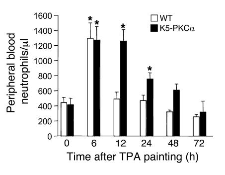
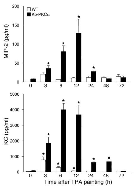
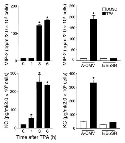
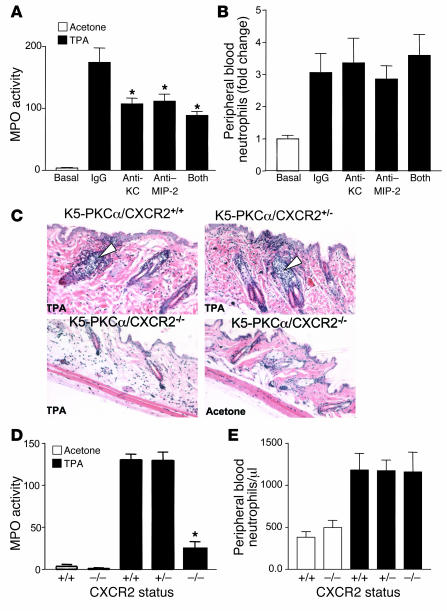
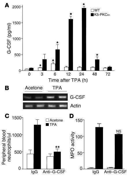
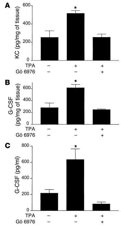
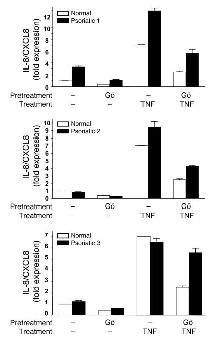
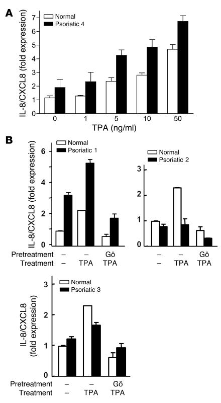
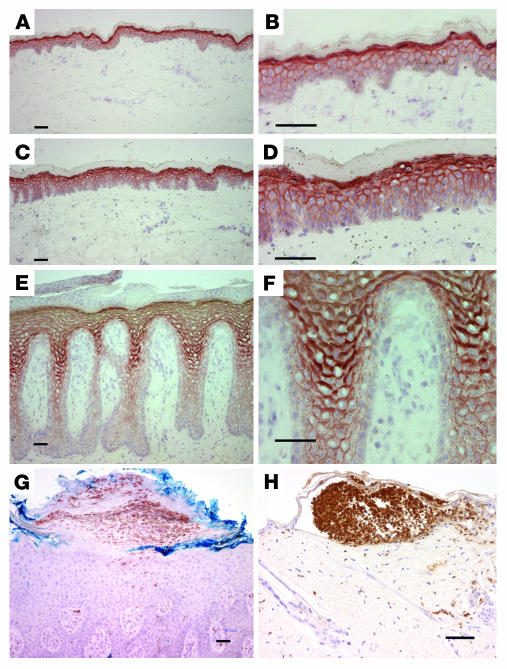
Transgenic mice overexpressing PKCalpha in the epidermis (K5-PKCalpha mice) exhibit an inducible severe intraepidermal neutrophilic inflammation and systemic neutrophilia when PKCalpha is activated by topical 12-O-tetradecanoylphorbol-13-acetate (TPA). This inducible model of cutaneous inflammation was used to define mediators of skin inflammation that may have clinical relevance. Activation of cutaneous PKCalpha increased the production of the chemotactic factors cytokine-induced neutrophil chemoattractant (KC) and macrophage inflammatory protein 2 (MIP-2) in murine plasma. TPA treatment of cultured K5-PKCalpha keratinocytes also released KC and MIP-2 into culture supernatants through an NF-kappaB-dependent pathway. MIP-2 and KC mediated the infiltration of neutrophils into the epidermis, since this was prevented by ablating CXCR2 in K5-PKCalpha mice or administering neutralizing antibodies against KC or MIP-2. The neutrophilia resulted from PKCalpha-mediated upregulation of cutaneous G-CSF released into the plasma independent of CXCR2. These responses could be inhibited by topical treatment with a PKCalpha-selective inhibitor. Inhibiting PKCalpha also reduced the basal and TNF-alpha- or TPA-induced expression of CXCL8 in cultured psoriatic keratinocytes, suggesting that PKCalpha activity may contribute to psoriatic inflammation. Thus, skin can be the source of circulating factors that have both local and systemic consequences, and these factors, their receptors, and possibly PKCalpha could be therapeutic targets for inhibition of cutaneous inflammation.


Figures

Figure 1
Figure 1. K5-PKCα mice develop a sustained neutrophilia in response to TPA painting.
A single dose of TPA (1 μg) in acetone was applied to the shaved backs of K5-PKCα transgenic mice and WT littermates. Blood was drawn at various times, and differential wbc counts were performed. Bars represent the mean number of neutrophils per microliter of blood ± SEM for at least 7 mice per group for each time point. *P < 0.05 versus the respective control.
Figure 2
Figure 2. K5-PKCα mice have increased circulating levels of MIP-2 and KC in response to TPA painting.
A single dose of TPA (1 μg) in acetone was applied to the shaved backs of K5-PKCα transgenic and WT littermates, blood was drawn at various times, and ELISA for KC and MIP-2 was performed on sera. Bars represent the mean ± SEM for 6 animals, and results are representative of at least 2 independent experiments. *P < 0.05 versus the respective control.
Figure 3
Figure 3. K5-PKCα primary keratinocytes produce MIP-2 and KC through PKCα-mediated NF-κB activation.
Left panels: Culture supernatants from K5-PKCα primary keratinocytes were collected at 1, 3, and 6 hours after TPA treatment. KC and MIP-2 concentrations were determined by ELISA. Bars represent the mean ± SEM of triplicate determinations. Results are representative of 3 independent experiments. *P < 0.05 versus time 0. Right panels: KC and MIP-2 concentrations in culture supernatant collected from K5-PKCα keratinocytes transduced with A-CMV (control) or degradation-resistant IκBα (IκBαSR) adenovirus after a 3-hour TPA or DMSO treatment. Bars represent the mean ± SEM of triplicate determinations. Results are representative of 3 independent experiments. *P < 0.05 versus the respective DMSO-treated/A-CMV control.
Figure 4
Figure 4. Blocking CXCR2 ligands or CXCR2 deficiency prevents neutrophil infiltration in the skin of K5-PKCα mice.
A single dose of TPA (1 μg) in acetone was applied to the shaved backs of K5-PKCα mice that had been injected intravenously with either control IgG, KC-neutralizing antibodies, MIP-2–neutralizing antibodies, or a combination of KC- and MIP-2–neutralizing antibodies. Skin was collected 6 hours later, and MPO activity was determined (A). Blood was collected at sacrifice, and the number of peripheral blood neutrophils was determined from differential wbc counts (B). For A and B, bars represent the mean ± SEM for 4 animals, and results are representative of 4 experiments. *P < 0.05 versus IgG control group. (C) K5-PKCα mice expressing CXCR2 (K5-PKCα/CXCR2+/+), K5-PKCα mice heterozygous for CXCR2 (K5-PKCα/CXCR2+/–), and K5-PKCα mice deficient for CXCR2 (K5-PKCα/CXCR2–/–) were TPA painted; skin was collected 6 hours later, and sections were stained with H&E. White arrowheads show early stage of neutrophil infiltration into the hair follicles. MPO was determined in skin extracts from the mouse groups depicted in C (D) as well as peripheral blood neutrophil counts (E). For D and E, bars represent the mean ± SEM for 7 animals, and results are representative of 2 experiments. *P < 0.05 versus TPA-treated K5-PKCα/CXCR2+/+ group.
Figure 5
Figure 5. Blocking G-CSF prevents neutrophilia in K5-PKCα mice.
(A) A single dose of TPA (1 μg) in acetone was applied to the shaved backs of K5-PKCα transgenic mice and WT littermates. Blood was drawn at various times, and ELISA for G-CSF was performed on sera. Bars represent the mean ± SEM for 6 animals, and results are representative of at least 2 independent experiments. *P < 0.05 versus the respective control. (B) Agarose gel stained with ethidium bromide showing RT-PCR product for G-CSF and actin. RNA was extracted from K5-PKCα skin punch biopsy samples 3 hours after TPA or acetone treatment. Each lane represents an individual mouse. Peripheral blood neutrophil counts (C) and skin MPO activity (D) 6 hours after a single dose of TPA (1 μg) in acetone was applied to the shaved backs of K5-PKCα mice that had been injected intraperitoneally with either control IgG or G-CSF–neutralizing antibodies. Bars represent the mean ± SEM for 8 animals, and results are representative of 2 independent experiments. **P < 0.0001 versus TPA-treated IgG group. NS, not significant versus TPA-treated IgG group.
Figure 6
Figure 6. Prevention of KC and G-CSF expression by the classical PKC inhibitor Gö 6976.
Gö 6976 was applied once topically just prior to TPA treatment. KC and G-CSF expression was quantified in skin extract (A and B, respectively), and levels circulating G-CSF (C) were determined in serum. The control group received the same volume of the solvent. Bars represent the mean ± SEM for 4 animals, and results are representative of 2 independent experiments. *P < 0.05 versus no TPA treatment.
Figure 7
Figure 7. PKCα inhibition attenuates abnormal levels of basal and induced CXCL8 in keratinocytes from psoriatic patients.
Real-time PCR analysis of CXCL8 mRNA expression in normal and psoriatic keratinocytes from lesional skin biopsy samples from psoriatic patients (Psoriatic 1–3) or normal skin of healthy controls treated as described in Methods. “Normal” is representative of cultured keratinocytes from 3 healthy controls. Bars represent the mean value of triplicate determinations from a single patient. Results are expressed as fold increase in expression compared with that in untreated normal keratinocytes.
Figure 8
Figure 8. Keratinocytes from a subset of psoriatic patients express higher CXCL8 mRNA levels in response to TPA.
(A) Real-time PCR analysis of CXCL8 mRNA expression in keratinocytes cultured from lesional skin biopsy samples from a psoriatic patient or skin of a healthy control treated with increasing doses of TPA. (B) CXCL8 mRNA expression in patient and control keratinocyte cultures shown in Figure 7. Cultures were stimulated for 1 hour with TPA (5 ng/ml) with or without incubation with 5 μM Gö 6976 for the previous 30 minutes (B). “Normal” is representative of cultured keratinocytes from 3 healthy controls. Bars represent mean value of triplicate determinations from a single patient. Results are expressed as fold increase in expression compared with that in untreated normal keratinocytes.
Figure 9
Figure 9. Prominent PKCα expression in the epidermis of psoriatic patients.
Frozen sections from normal (A and B), nonlesional (C and D), and psoriatic (E and F) epidermis were stained with monoclonal antibodies to PKCα and detected by immunoperoxidase. Results are typical of samples from 4 patients. Representative example of immunostaining for neutrophils in pustular psoriasis epidermis (G) and TPA-treated K5-PKCα mouse epidermis (H). PKCα staining in G (data not shown) was identical to E and F. Scale bars, 50 μm.

Similar articles

Cited by

References

    1. Kupper T.S., Fuhlbrigge R.C. Immune surveillance in the skin: mechanisms and clinical consequences. Nat. Rev. Immunol. 2004;4:211–222. - PMC - PubMed
    1. Murphy P.M., et al. International union of pharmacology. XXII. Nomenclature for chemokine receptors. Pharmacol. Rev. 2000;52:145–176. - PubMed
    1. Murphy P.M. International Union of Pharmacology. XXX. Update on chemokine receptor nomenclature. Pharmacol. Rev. 2002;54:227–229. - PubMed
    1. Zlotnik A., Yoshie O. Chemokines: a new classification system and their role in immunity. Immunity. 2000;12:121–127. - PubMed
    1. Bozic C.R., et al. Expression and biologic characterization of the murine chemokine KC. . J. Immunol. 1995;154:6048–6057. - PubMed

Publication types

MeSH terms