

64-element intraluminal ultrasound cylindrical phased array for transesophageal thermal ablation under fast MR temperature mapping: an ex vivo study
- PMID: 16964871
- PMCID: PMC1890449
- DOI: 10.1118/1.2218064
64-element intraluminal ultrasound cylindrical phased array for transesophageal thermal ablation under fast MR temperature mapping: an ex vivo study
Abstract
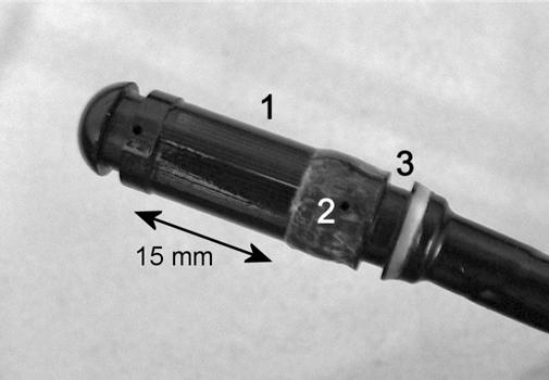
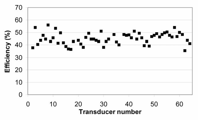
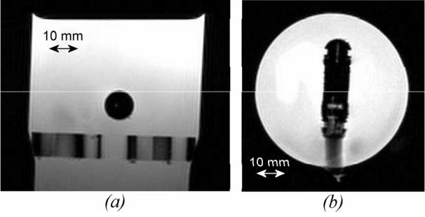
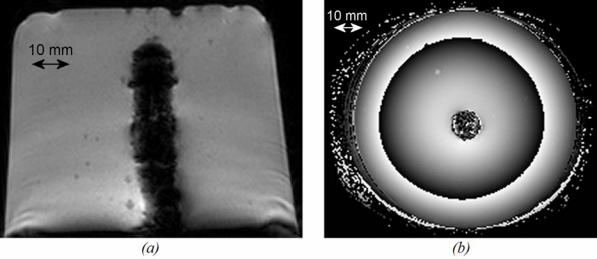
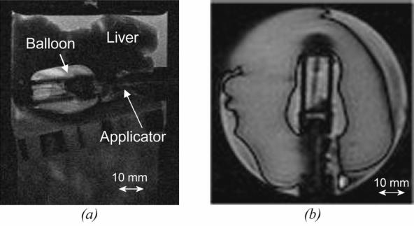
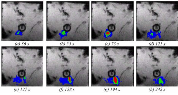
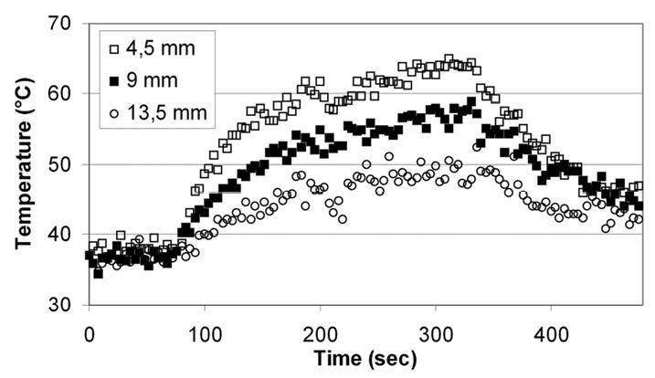
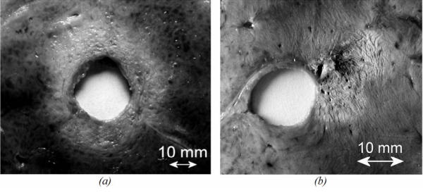
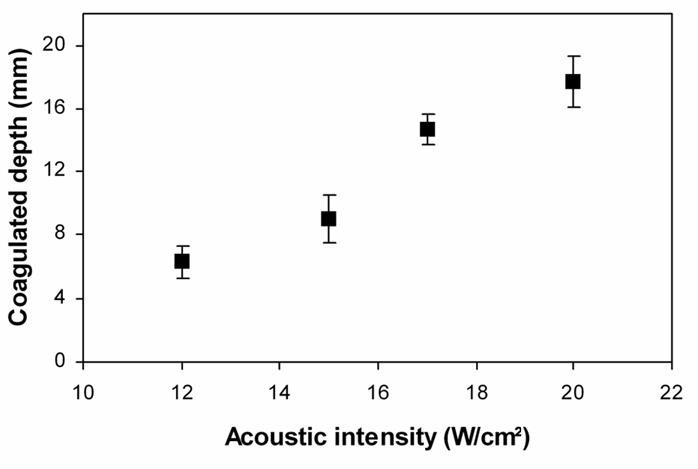
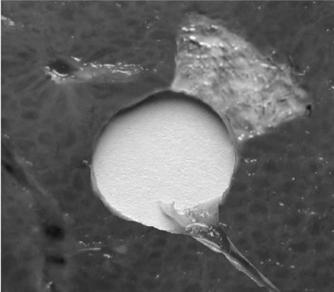
This work was undertaken to investigate the feasibility of using a cylindrical phased array for transoesophaeal thermal ablation under magnetic resonance (MR) imaging guidance. Sixty-four transducers (0.45 mm wide by 15 mm tall), operating at 4.6 MHz, were spread around the periphery of a 10.6-mm-diam cylinder. The head of the applicator was covered with a 65-microm thick latex balloon attached using watertight seals. This envelope was inflated with degassed water to provide acoustic coupling between the transducer and the tissues. The underlying operating principle of this applicator is to rotate a plane ultrasound beam electronically. For this purpose, eight adjacent transducers were excited with appropriate delay times so as to generate a plane wave. The exposure direction was changed by exciting a different set of eight elements. Ex vivo experiments conducted on 47 samples of pig liver under MR temperature monitoring demonstrated the ability of this applicator to generate cylindrical or sector-based coagulation necroses at depths up to 19 mm with excellent angular precision by applying 20 W/cm2. MR thermometry was performed in "real-time" with segmented echo-planar imaging gradient echo sequences. The temporal resolution was approximately 3 s/ image. The average value for the temperature baseline in liver tissue close to the applicator was 0.3 degrees C (+/- 0.6 degrees C). The thermal dose delivered in tissues was computed on-line during temperature imaging. Excellent MR compatibility was demonstrated, all MR acquisitions were performed without susceptibility artifacts or radio-frequency interferences with the ultrasound device. Thermal lesions identified on post-treatment follow up showed good correlation with online MR thermometry data. The individual differences between measurements performed visually and using MRI thermal dose maps were about 11% of volume. This study demonstrated the feasibility of thermal ablation using a phased array intraluminal ultrasound applicator and on-line MR monitoring.
Figures


Similar articles
-
Ultrasound cylindrical phased array for transoesophageal thermal therapy: initial studies.Phys Med Biol. 2002 Dec 7;47(23):4191-203. doi: 10.1088/0031-9155/47/23/306. Phys Med Biol. 2002. PMID: 12502043
-
Endocavitary thermal therapy by MRI-guided phased-array contact ultrasound: experimental and numerical studies on the multi-input single-output PID temperature controller's convergence and stability.Med Phys. 2009 Oct;36(10):4726-41. doi: 10.1118/1.3215534. Med Phys. 2009. PMID: 19928104
-
Endoluminal ultrasound applicators for MR-guided thermal ablation of pancreatic tumors: Preliminary design and evaluation in a porcine pancreas model.Med Phys. 2016 Jul;43(7):4184. doi: 10.1118/1.4953632. Med Phys. 2016. PMID: 27370138 Free PMC article.
-
Catheter-based ultrasound applicators for selective thermal ablation: progress towards MRI-guided applications in prostate.Int J Hyperthermia. 2004 Nov;20(7):739-56. doi: 10.1080/02656730410001721816. Int J Hyperthermia. 2004. PMID: 15675669 Review.
-
Technical advances in motion-robust MR thermometry.Magn Reson Med. 2024 Jul;92(1):15-27. doi: 10.1002/mrm.30057. Epub 2024 Mar 19. Magn Reson Med. 2024. PMID: 38501903 Free PMC article. Review.
Cited by
-
Deployable ultrasound applicators for endoluminal delivery of volumetric hyperthermia.Int J Hyperthermia. 2021 Aug 10;38(1):1188-1204. doi: 10.1080/02656736.2021.1936216. Int J Hyperthermia. 2021. PMID: 34376103 Free PMC article.
-
Catheter-based ultrasound technology for image-guided thermal therapy: current technology and applications.Int J Hyperthermia. 2015 Mar;31(2):203-15. doi: 10.3109/02656736.2015.1006269. Epub 2015 Mar 23. Int J Hyperthermia. 2015. PMID: 25799287 Free PMC article. Review.
-
Treatment of rabbit liver cancer in vivo using miniaturized image-ablate ultrasound arrays.Ultrasound Med Biol. 2011 Oct;37(10):1609-21. doi: 10.1016/j.ultrasmedbio.2011.05.850. Epub 2011 Aug 6. Ultrasound Med Biol. 2011. PMID: 21821349 Free PMC article.
-
Thermal therapy of pancreatic tumours using endoluminal ultrasound: Parametric and patient-specific modelling.Int J Hyperthermia. 2016;32(2):97-111. doi: 10.3109/02656736.2015.1119892. Epub 2016 Jan 21. Int J Hyperthermia. 2016. PMID: 27097663 Free PMC article.
-
Deployable cylindrical phased-array applicator mimicking a concentric-ring configuration for minimally-invasive delivery of therapeutic ultrasound.Phys Med Biol. 2019 Jun 10;64(12):125001. doi: 10.1088/1361-6560/ab2318. Phys Med Biol. 2019. PMID: 31108478 Free PMC article.
References
-
- Inoue H, Tani M, Nagai K, Kawano T, Takeshita K, Endo M, Iwai T. Treatment of esophageal and gastric tumors. Endoscopy. 1999;31:47–55. - PubMed
-
- Coia LR, Minsky BD, Berkey BA, John MJ, Haller D, Landry J, Pisansky TM, Willett CG, Hoffman JP, Owen JB, Hanks GE. Outcome of patients receiving radiation for cancer of the esophagus: results of the 1992–1994 Patterns of Care Study. J Clin Oncol. 2000;18:455–462. - PubMed
-
- De Palma GD, di Matteo E, Romano G, Fimmano A, Rondinone G, Catanzano C. Plastic prosthesis versus expandable metal stents for palliation of inoperable esophageal thoracic carcinoma: a controlled prospective study. Gastrointest Endosc. 1996;43:478–482. - PubMed
-
- Konigsrainer A, Riedmann B, De Vries A, Ofner D, Spechtenhauser B, Aigner F, Fritsch E, Margreiter R. Expandable metal stents versus laser combined with radiotherapy for palliation of unresectable esophageal cancer: a prospective randomized trial. Hepatogastroenterology. 2000;47:724–727. - PubMed
-
- Gelet A, Chapelon JY, Poissonnier L, Bouvier R, Rouviere O, Curiel L, Janier M, Vallancien G. Local recurrence of prostate cancer after external beam radiotherapy: early experience of salvage therapy using high-intensity focused ultrasonography. Urology. 2004;63:625–629. - PubMed
Publication types
MeSH terms
LinkOut - more resources
Full Text Sources
Medical
