Review
. 2006 Oct;19(5):380-94.
doi: 10.1111/j.1600-0749.2006.00318.x.

The other pigment cell: specification and development of the pigmented epithelium of the vertebrate eye

Affiliations
Review

The other pigment cell: specification and development of the pigmented epithelium of the vertebrate eye

Kapil Bharti et al. Pigment Cell Res. 2006 Oct.

Abstract

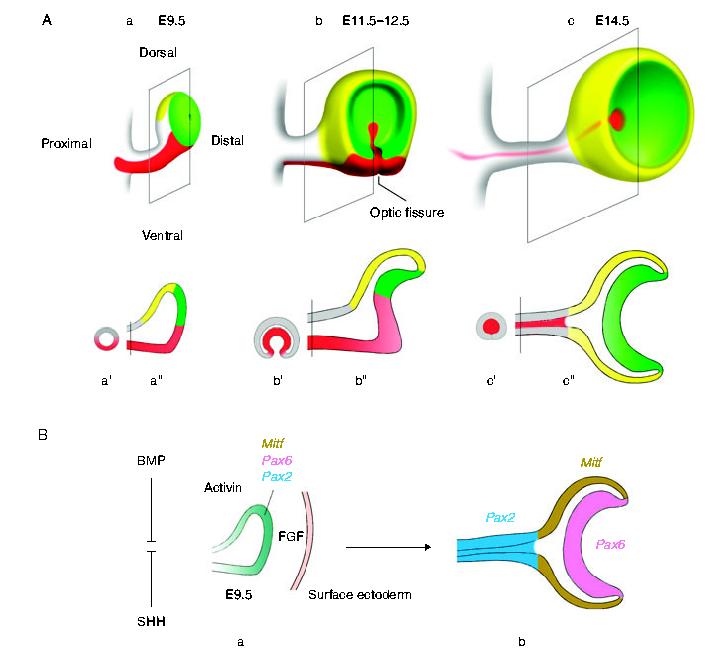
Vertebrate retinal pigment epithelium (RPE) cells are derived from the multipotent optic neuroepithelium, develop in close proximity to the retina, and are indispensible for eye organogenesis and vision. Recent advances in our understanding of RPE development provide evidence for how critical signaling factors operating in dorso-ventral and distal-proximal gradients interact with key transcription factors to specify three distinct domains in the budding optic neuroepithelium: the distal future retina, the proximal future optic stalk/optic nerve, and the dorsal future RPE. Concomitantly with domain specification, the eye primordium progresses from a vesicle to a cup, RPE pigmentation extends towards the ventral side, and the future ciliary body and iris form from the margin zone between RPE and retina. While much has been learned about the molecular networks controlling RPE cell specification, key questions concerning the cell proliferative parameters in RPE and the subsequent morphogenetic events still need to be addressed in greater detail.


Figures

Figure 1
Figure 1
The development of the mouse eye and its territorialization. (A) Overview/fate map: (a-c) Eye development in the mouse starts at around embryonic day (E) 7.5 from an ‘eye field’ of cells in the midline of the neural plate at the level of the prospective diencephalon. This portion of the neuroepithelium evaginates laterally in a dorsal-distal direction, extending towards the surface ectoderm (which is omitted in the figure for clarity). The process generates proximally the optic stalk (which will become the optic nerve) and distally the optic vesicle (which will become neuroretina, RPE, ciliary body, and iris). (a) The optic vesicle is readily visible at E9.5. (b) When the vesicle contacts the surface ectoderm, its distal-most part becomes indented to form the optic cup. The indentation extends towards the ventral part of the optic stalk, generating a fissure along the ventral side of stalk and retina, called the optic fissure. The optic cup is now a highly polarized structure, both in the dorso-ventral and proximal-distal orientation. (c) The fissure progressively seals and is usually closed by E13.5. As a result of this process, the ventral optic stalk epithelium comes to lie inside the stalk where it will generate the astrocytes of the optic nerve. With the closure of the fissure, the cup’s outer wall is entirely composed of RPE. This morphogenetic process is further schematically illustrated in the cross-sections through the optic stalk (a’-c’) and the coronal sections through the optic vesicle/optic cup (a”-c”). Not shown in these drawings is the process of lens formation from the overlying surface ectoderm. Color code for (A): yellow, future RPE; green, future retina; red: ventral optic stalk and derivatives; gray (in sections): dorsal optic stalk and derivatives. (B) Gene expression during territorialization: The budding optic vesicle expresses many factors that initially overlap (a), but eventually become responsible for distinct eye tissues (b). This requires sorting out their expression patterns into the different domains of the neuroepithelium, a process that is controlled by cell-extrinsic factors such as activins and FGFs that emanate from specific local points and locally impinge on gene expression in the nearby neuroepithelium (see text). For instance, the overlapping expression patterns of MITF, PAX2, and Pax6 at E9.5 (a) are sorted into the expression domains indicated in (b) (see also Bäumer et al., 2003). PAX2, for instance, becomes concentrated in the optic stalk as early as E10.5 and eventually is found in the astrocytes of the optic nerve along with Vax1 (not shown, see text). Equally early, MITF is downregulated in the future retina while Pax6 stays on. In contrast, in the future RPE, where MITF becomes prominent, Pax6 fades away, though only several days later. Hence, the drawing on the right (b) represents not a specific developmental time point but a conceptualization of a dynamic process. Color code for (B): blue-green: overlapping expression of MITF, PAX6, and PAX2 during early vesicle formation. Later stages: colors as indicated in the figure.
Figure 2
Figure 2
The role of Mitf in RPE formation. MITF is a basic-helix-loop-helix-leucine zipper transcription factor expressed in the developing RPE. (A and B) Cross-section through the dorsal RPE of an Mitf+/+, albino mouse embryo at E14.5 and its counterpart carrying an Mitf allele encoding a protein that lacks part of the basic domain (Mitf?mi-ew). both embryo sections were stained with a rabbit anti-MITF antiserum. Brackets mark the RPE monolayer in (A) and the thickened RPE in (B). Note that in the mutant (B), the part that is going to become the ’transdifferentiated’ second retina (arrow) no longer expresses MITF protein. This second retina soon degenerates after birth, leading to additional abnormalities in the adjacent original retina. (C-E) Sections through the posterior RPE and adjacent retina in 6-week-old wild type (pigmented mouse) and two Mitf mutant compound heterozygotes (for allele description, see Arnheiter et al., 2002; Steingrimsson et al., 2004). In the wild type (C), one normally sees a pigmented RPE sandwiched between the rod outer segments (ros) of the retina and the pigmented choroid. (D) In mice carrying a relatively mild Mitf allele, Mitfmi-vit, here in combination with a null allele (Mitfmi-vga-9), individual RPE cells are unpigmented (arrows), while others retain pigment (arrowheads). The choroid, however, is entirely unpigmented. In these mice, the retina progressively degenerates. (E) In mice carrying a more severe Mitf mutation (MitfMi-or/mi-vga-9), there is considerable hypercellularity in an RPE that remains totally unpigmented. The adjacent retina is abnormal and lacks lamination.
Figure 3
Figure 3
FGF downregulates Mitf at the mRNA level. The frontal part of the heads of E9.5 mouse embryos were explanted. A polyacrylamide bead, coated with bovine serum albumin and labeled ‘control bead’, was implanted on one side near the optic vesicle. Another bead, coated with FGF2 and labeled ‘FGF2 bead’, was implanted close to the optic vesicle on the other side. After 72 h, the heads were fixed and processed for whole mount non-radioactive in situ hybridization using an Mitf RNA probe. Note that on the control side, Mitf mRNA is expressed as expected (dark brown stain) but no Mitf label is seen on the FGF2 treated side.
Figure 4
Figure 4
Ciliary body and iris formation. The ciliary body and the iris form as a special elongation from the rim of the optic cup, called the ciliary margin zone or CMZ, where the outer layer (RPE) turns around into the inner layer (retina). The picture on the left shows a section through an albino eye of an Mitf+/+ mouse embryo at E16.5. This section was labeled by non-radioactive in situ hybridization for a pigmentation gene, Dct. Note the presence of Dct-positive neural crest-derived cells at the surface and close to the CMZ. The two pictures on the right show sections through adult eyes from wild type and a compound heterozygous Mitf mutant, Mitfmi-bw/mi-vga-9. The CMZ has given rise to the ciliary processes which provide the excess tissue needed during closure of the pupil. The extension of the retina forms the unpigmented ciliary epithelium and the extension of the RPE the pigmented back of the iris. The front of the iris is populated with neural crest-derived melanocytes. The distinct origin of these two pigmented layers of the iris can be seen in the mutant eye in which the generation of neural crest-derived melanocytes but not of RPE cells is affected. The irises of such mutant mice have a pigmented layer on the back but lack pigmented cells in the front; here, only unpigmented stromal cells are found. Nevertheless, on visual inspection, the mutant eyes are black, in contrast to the red eyes of albino animals which lack melanin in both layers. Hence, pigmentation of the RPE and the back layer of the iris is sufficient for the visual appearance of a black eye.

Similar articles

Cited by

References

    1. Ahmad I, Tang L, Pham H. Identification of neural progenitors in the adult mammalian eye. Biochem. Biophys. Res. Commun. 2000;270:517–521. - PubMed
    1. Amato MA, Boy S, Perron M. Hedgehog signaling in vertebrate eye development: a growing puzzle. Cell Mol. Life Sci. 2004;61:899–910. - PMC - PubMed
    1. Arnheiter H. Evolutionary biology. Eyes viewed from the skin. Nature. 1998;391:632–633. - PubMed
    1. Arnheiter H, Hou L, Nguyen MT, Nakayama A, Champagne B, Hallsson JH, Bismuth K. The role of microphthalmia in pigment cell development. In: Ortonne J.p., Ballotti R., editors. Mechanisms of Suntanning. Martin Dunitz Publication; London: 2002. pp. 49–63.
    1. Arnheiter H, Hou L, Nguyen MTT, Bismuth K, Csermely T, Murakami H, Skuntz S, Liu W, Bharti K. Mitf—A matter of life and death for the developing melanocyte. In: Hearing V, Leong SPL, editors. From Melanocytes to Malignant Melanoma. Humana Press; Totowa, NJ: 2006. pp. 27–49.

Publication types