. 2006 Nov;97(11):1182-90.
doi: 10.1111/j.1349-7006.2006.00322.x. Epub 2006 Sep 12.

High expression of insulin-like growth factor binding protein-3 is correlated with lower portal invasion and better prognosis in human hepatocellular carcinoma

Affiliations

High expression of insulin-like growth factor binding protein-3 is correlated with lower portal invasion and better prognosis in human hepatocellular carcinoma

Shinichi Aishima et al. Cancer Sci. 2006 Nov.

Abstract

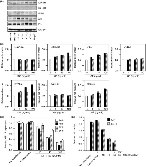
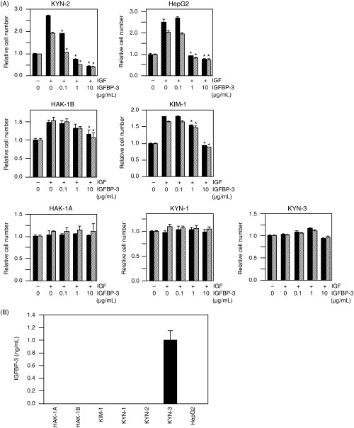
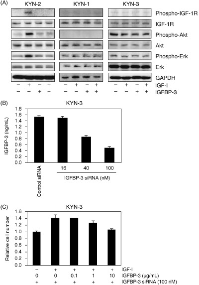
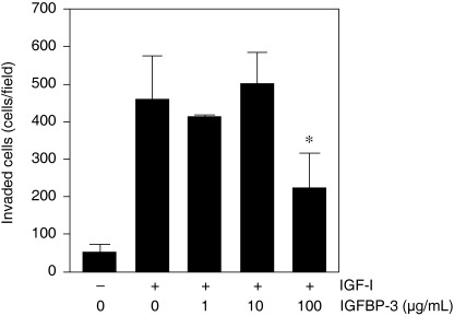
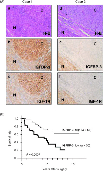
Insulin-like growth factor binding protein-3 (IGFBP-3) modulates cell proliferation of various cancer cell types. However, it remains unclear how IGF-IGFBP-3-signaling is involved in growth and progression of hepatocellular carcinoma (HCC). The aim of the present study was to evaluate the role of IGFBP-3 in HCC. Type 1 receptor for IGF (IGF-1R) was expressed at various levels in the seven lines examined, but IGF-2R was not expressed. Of the seven lines, the growth of HAK-1B, KIM-1, KYN-2 and HepG2 cells was stimulated in a dose-dependent manner by the exogenous addition of IGF-I or IGF-II, but the HAK-1A, KYN-1 and KYN-3 cell lines showed no growth. Exogenous addition of IGFBP-3 markedly blocked IGF-I and IGF-II-stimulated cell growth of KYN-2 and HepG2 cells, and moderately stimulated that of KIM-1 and HAK-1B cells, but no growth of the KYN-1, KYN-3 and HAK-1A cell lines was observed. IGF-I enhanced the phosphorylation of IGF-1R, Akt and Erk1/2 in KYN-2 cells, and coadministration of IGFBP-3 blocked all types of activation by IGF-I investigated here. In contrast, no such activation by IGF-I was detected in KYN-3 cells. IGFBP-3 also suppressed IGF-I-induced cell invasion by KYN-2 cells. Moreover, we were able to observe the apparent expression of IGFBP-3 in KYN-3 cells, but not in the other six cell lines. Furthermore reduced expression of IGFBP-3, but not that of IGF-1R, was significantly correlated with tumor size, histological differentiation, capsular invasion and portal venous invasion. Low expression of IGFBP-3 was independently associated with poor survival. IGFBP-3 could be a molecular target of intrinsic importance for further development of novel therapeutic strategy against HCC.


Figures

Figure 1
Figure 1
(A) Expression of type 1 insulin‐like growth factor (IGF) receptor (IGF‐1R), IGF‐2R, IRS‐1, Akt and Erk was determined by immunoblotting conducted on protein lysates extracted from these cell lines. The detection of glyceraldehyde‐3‐phosphate dehydrogenase (GAPDH) served as a loading control. (B) Effects of IGF‐I and IGF‐II on the proliferation of seven hepatocellular carcinoma cell lines. The cells were treated with or without IGF at concentrations of 1, 10 or 100 ng/mL for 72 h in serum‐free media, and then colorimetric WST assays were carried out. Each bar represents IGF‐I (closed bar) or IGF‐II (hatched bar). The data are expressed as the mean ± SD. (C) Inhibition by IGF‐1R small interfering RNA (siRNA) treatment of IGF‐1R gene expression of KYN‐2 cells. KYN‐2 cells were transfected with IGF‐1R siRNA at concentrations of 0, 16, 40 and 100 nM, and the cells were incubated for the periods of time indicated. After incubation, total RNA was extracted and gene silencing was analyzed by real‐time quantitative polymerase chain reaction. (D) Effect of IGF‐1R siRNA on the proliferation of IGF‐I (closed bar) or IGF‐II (hatched bar)‐stimulated KYN‐2 cells. The cells were stimulated with 100 ng/mL of IGF‐I or IGF‐II after 24 h of siRNA treatment, and a WST assay was carried out 72 h after IGF stimulation. The data are expressed as the mean ± SD.
Figure 2
Figure 2
(A) The effects of insulin‐like growth factor (IGF) binding protein (IGFBP)‐3 on IGF‐I‐ or IGF‐II‐dependent cell proliferation of seven hepatocellular carcinoma cell lines. The cells were incubated with either serum‐free medium, 100 ng/mL of IGF‐I (closed bar) or 100 ng/mL of IGF‐II (hatched bar) in the presence of various concentrations of IGFBP‐3 for 72 h. After incubation, colorimetric WST assays were carried out. *Significant differences (P < 0.01) compared with treatment with IGF alone in the absence of IGFBP‐3. The data are expressed as the mean ± SD. (B) Cellular production of IGFBP‐3 in KYN‐3 cells. IGFBP‐3 protein levels in the culture medium with the seven cell lines examined here were assayed quantitatively with enzyme‐linked immunosorbent assay systems. The data are the average of triplicate wells ± SD.
Figure 3
Figure 3
(A) Effects of insulin‐like growth factor (IGF)‐I and IGF binding protein (IGFBP)‐3 on the phosphorylation of type 1 IGF receptor (IGF‐1R), Akt and Erk in KYN‐2, KYN‐1 and KYN‐3 cells. Serum‐deprived cells were treated with 100 ng/mL IGF‐I and/or with 10 µg/mL IGFBP‐3 for 10 min. Cell lysates were blotted with the antibodies indicated. The detection of glyceraldehyde‐3‐phosphate dehydrogenase (GAPDH) served as a loading control. (B) Inhibition by IGFBP‐3 small interfering RNA (siRNA) treatment of IGFBP‐3 expression in KYN‐3 cells. KYN‐3 cells were transfected with IGFBP‐3 siRNA at concentrations of 0, 16, 40 and 100 nM, and the cells were incubated for 24 h. After incubation, IGFBP‐3 protein levels in the culture medium were assayed quantitatively with enzyme‐linked immunosorbent assay systems. The data are the average of triplicate wells ± SD. (C) Restoration of IGF‐I‐stimulated proliferation of KYN‐3 cells by IGFBP‐3 siRNA. KYN‐3 cells were treated for 24 h with 100 nM IGFBP‐3 siRNA and further incubation with 100 ng/mL of IGF‐I in the absence or presence of various doses of IGFBP‐3 for 72 h. The data are expressed as the mean ± SD.
Figure 4
Figure 4
Effect of insulin‐like growth factor (IGF) binding protein (IGFBP)‐3 on invasion by IGF‐stimulated KYN‐2 cells in Matrigel invasion assay. Serum‐deprived KYN‐2 cells (1 × 105) were seeded onto Matrigel‐coated filters in the upper chambers, and Dulbecco's modified Eagle's medium with or without IGF‐I (1000 ng/mL) in the presence of various concentrations of IGFBP‐3. The cell invasiveness was quantified as the mean cell number in five fields of view per filter. Columns, mean of three independent experiments; bars, ±SD.
Figure 5
Figure 5
(A) Immunohistochemical expression of insulin‐like growth factor (IGF) binding protein (IGFBP)‐3 in hepatocellular carcinoma (HCC). Two representative cases of well‐differentiated HCC (case 1) and poorly differentiated HCC (case 2). The expression of both IGFBP‐3 and type 1 IGF receptor (IGF‐1R) was stronger in the HCC than in the adjacent liver in the cases of well‐differentiated HCC (b,c). In contrast, cases of poorly differentiated HCC showed reduced expression of IGFBP‐3 and IGF‐1R in the HCC tissue compared with that of the adjacent liver tissue (e,f). (a,d) Hematoxylin–eosin (H–E) staining. C, cancerous region; N, non‐cancerous region. (B) The overall survival of patients with low IGFBP‐3 expression was significantly worse than that of patients with high IGFBP‐3 expression (P = 0.0007).
Figure 6
Figure 6
Correlation between hepatocellular carcinoma (HCC) staging and serum insulin‐like growth factor (IGF)‐I levels or serum IGF binding protein (IGFBP)‐3 levels in patients with HCC (n = 92). (A) Correlation between HCC staging and serum IGF‐I levels. (B) correlation between HCC staging and serum IGFBP‐3 levels. Patients of stage I (n = 22), II (n = 17), III (n = 31) and IV (n = 22).

References

    1. Shimada M, Takenaka K, Gion T et al. Prognosis of recurrent hepatocellular carcinoma: a 10‐year surgical experience in Japan. Gastroenterology 1996; 111: 720–6. - PubMed
    1. Adachi E, Maehara S, Tsujita E et al. Clinicopathologic risk factors for recurrence after a curative hepatic resection for hepatocellular carcinoma. Surgery 2002; 131: 148–52. - PubMed
    1. Scharf JG, Schmidt‐Sandte W, Pahernik SA, Ramadori G, Braulke T, Hartmann H. Characterization of the insulin‐like growth factor axis in a human hepatoma cell line (PLC). Carcinogenesis 1998; 19: 2121–8. - PubMed
    1. LeRoith D, Werner H, Beitner‐Johnson D, Roberts CT Jr. Molecular and cellular aspects of the insulin‐like growth factor I receptor. Endocr Rev 1995; 16: 143–63. - PubMed
    1. Ito T, Sasaki Y, Wands JR. Overexpression of human insulin receptor substrate 1 induces cellular transformation with activation of mitogen‐activated protein kinases. Mol Cell Biol 1996; 16: 943–51. - PMC - PubMed

Publication types

MeSH terms

Substances