

Tissue- and hormone-dependent progesterone receptor distribution in the rat uterus
- PMID: 16965620
- PMCID: PMC1586009
- DOI: 10.1186/1477-7827-4-47
Tissue- and hormone-dependent progesterone receptor distribution in the rat uterus
Abstract
Background: Estradiol (E2) and progesterone (P) are well known regulators of progesterone receptor (PR) expression in the rat uterus. However, it is not known which receptor subtypes are involved. Little knowledge exist about possible differences in PR regulation through ERalpha or ERbeta, and whether the PR subtypes are differently regulated depending on ER type bound. Thus, in the present study PR immunostaining has been examined in uteri of ovariectomized (ovx) rats after different treatments of estrogen and P, in comparison with that in immature, cycling, and pregnant animals.
Methods: The uteri were collected from 1) ovx rats treated with E2 and/or P; 2) immature rats, intact cycling rats and animals pregnant day 8 and 18; 3) ovx rats treated with E2 or an estrogen receptor (ER)alpha agonist or an ERbeta agonist. Two antibodies were used, one detecting PRA+B and another one specific for PRB. Real-time PCR was used to determine mRNA levels for PRAB and PRB in experiment 3.
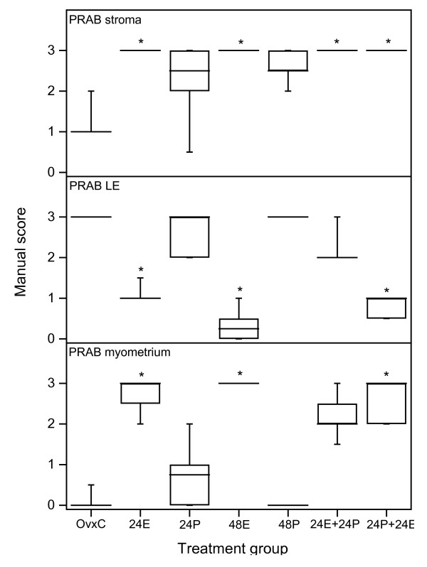
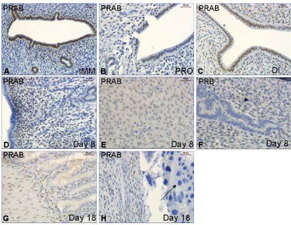
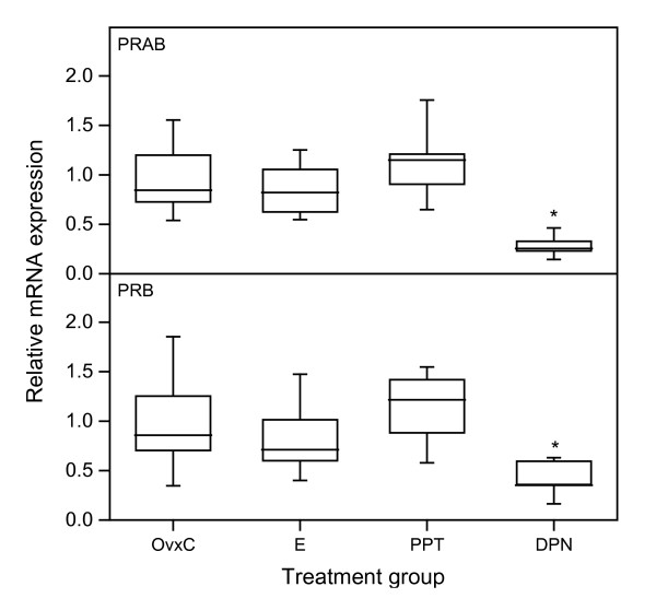
Results: In stroma and myometrium faint staining was detected in ovx controls (OvxC), whereas E2 treatment resulted in strong staining. In contrast to this, in luminal epithelium (LE) the staining was strong in the OvxC group, whereas E2 treatment during the last 24 hrs before sacrifice caused a decrease. Similar to OvxC the LE of the immature animals was strongly stained. In the pregnant rats LE was negative, well in agreement with the results seen after E2 treatment. In the pregnant animals the stroma and decidua was strongly stained for PRAB, but only faint for PRB, indicating that PRA is the most expressed isoform in this state. The increase in stromal and myometrial immunostaining after E2 treatment was also found after treatment with the ERalpha agonist PPT. The ERbeta agonist DPN caused a decrease of the PR mRNA levels, which was also found for PRAB and PRB immunostaining in the GE.
Conclusion: Stromal and myometrial PRAB levels are increased via ERalpha, as shown by treatment with E2 and the ERalpha agonist PPT, while the levels in LE are decreased. The uterine stroma of pregnant rats strongly expressed PRAB, but very little PRB, which is different to E2 treated ovx animals where both PRAB and PRB are strongly expressed. The ERbeta agonist DPN decreased the mRNA levels of PRAB and PRB, as well as the PRAB protein level in GE. These results suggest that ERbeta signals mainly down-regulate PR levels in the epithelial cells. ERalpha, on the other hand, up-regulates PR levels in the stroma and myometrium while it decreased them in LE. Thus, the effects from E2 and PPT on the mRNA levels, as determined by PCR, could be annihilated since they are increased and decreased depending on cell type. The distribution and amount of PR isoforms strongly depend on the hormonal milieu and cell type within the rat uterus.
Figures


Similar articles
-
The estrogen receptor alpha sigma3 mRNA splicing variant is differentially regulated by estrogen and progesterone in the rat uterus.J Endocrinol. 2005 Jul;186(1):51-60. doi: 10.1677/joe.1.06099. J Endocrinol. 2005. PMID: 16002535
-
Characterization of the pharmacologic profile of a standardized soy extract in the ovariectomized rat model of menopause: effects on bone, uterus, and lipid profile.Menopause. 2005 Sep-Oct;12(5):589-600. doi: 10.1097/01.GME.0000156348.61767.D5. Epub 2005 Sep 1. Menopause. 2005. PMID: 16145313
-
Regulation of hepatic progesterone and estrogen receptors in the female turtle, Chrysemys picta: relationship to vitellogenesis.Gen Comp Endocrinol. 2004 Apr;136(2):232-40. doi: 10.1016/j.ygcen.2003.12.016. Gen Comp Endocrinol. 2004. PMID: 15028527
-
Myometrial progesterone responsiveness and the control of human parturition.J Soc Gynecol Investig. 2004 May;11(4):193-202. doi: 10.1016/j.jsgi.2003.12.004. J Soc Gynecol Investig. 2004. PMID: 15120691 Review.
-
Progesterone and placental hormone actions on the uterus: insights from domestic animals.Biol Reprod. 2004 Jul;71(1):2-10. doi: 10.1095/biolreprod.103.024133. Epub 2004 Feb 18. Biol Reprod. 2004. PMID: 14973264 Review.
Cited by
-
Ovulation: Parallels With Inflammatory Processes.Endocr Rev. 2019 Apr 1;40(2):369-416. doi: 10.1210/er.2018-00075. Endocr Rev. 2019. PMID: 30496379 Free PMC article. Review.
-
Co-administering Melatonin With an Estradiol-Progesterone Menopausal Hormone Therapy Represses Mammary Cancer Development in a Mouse Model of HER2-Positive Breast Cancer.Front Oncol. 2019 Jul 9;9:525. doi: 10.3389/fonc.2019.00525. eCollection 2019. Front Oncol. 2019. PMID: 31355130 Free PMC article.
-
Progesterone distribution in the trigeminal system and its role to modulate sensory neurotransmission: influence of sex.J Headache Pain. 2023 Nov 14;24(1):154. doi: 10.1186/s10194-023-01687-x. J Headache Pain. 2023. PMID: 37957603 Free PMC article.
-
Inflammatory status influences aromatase and steroid receptor expression in endometriosis.Endocrinology. 2008 Mar;149(3):1190-204. doi: 10.1210/en.2007-0665. Epub 2007 Nov 29. Endocrinology. 2008. PMID: 18048499 Free PMC article.
-
Effects of hormonally active agents on steroid hormone receptor expression and cell proliferation in the myometrium of ovariectomized macaques.Toxicol Pathol. 2011 Apr;39(3):508-15. doi: 10.1177/0192623311401045. Epub 2011 Mar 16. Toxicol Pathol. 2011. PMID: 21411722 Free PMC article.
References
Publication types
MeSH terms
Substances
LinkOut - more resources
Full Text Sources
Research Materials
