. 2006 Dec 1;300(1):434-60.
doi: 10.1016/j.ydbio.2006.08.007. Epub 2006 Aug 10.

A genomic view of the sea urchin nervous system

Affiliations

A genomic view of the sea urchin nervous system

R D Burke et al. Dev Biol. .

Abstract

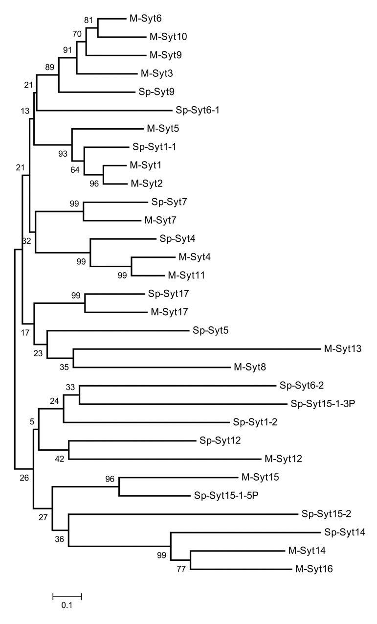
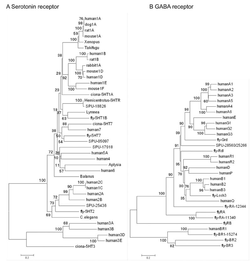
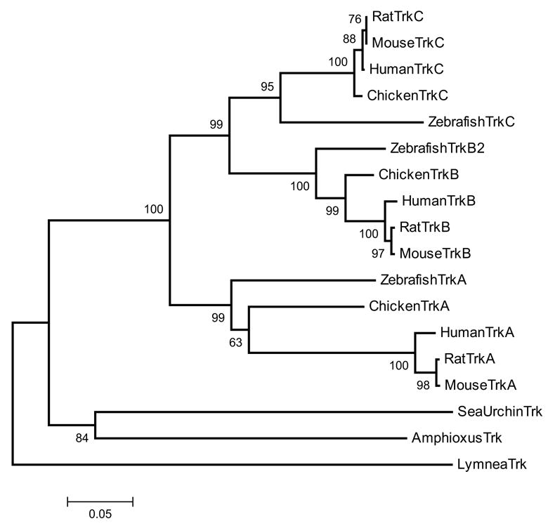
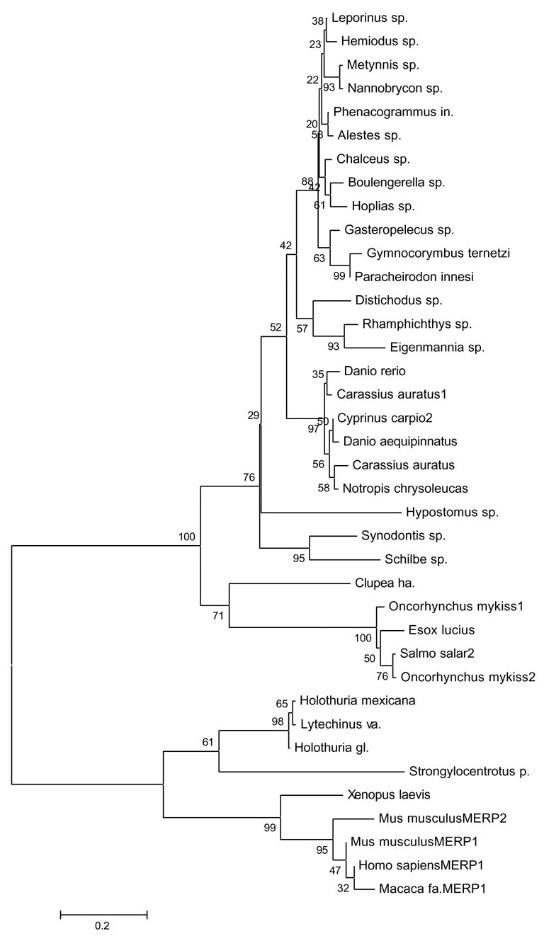
The sequencing of the Strongylocentrotus purpuratus genome provides a unique opportunity to investigate the function and evolution of neural genes. The neurobiology of sea urchins is of particular interest because they have a close phylogenetic relationship with chordates, yet a distinctive pentaradiate body plan and unusual neural organization. Orthologues of transcription factors that regulate neurogenesis in other animals have been identified and several are expressed in neurogenic domains before gastrulation indicating that they may operate near the top of a conserved neural gene regulatory network. A family of genes encoding voltage-gated ion channels is present but, surprisingly, genes encoding gap junction proteins (connexins and pannexins) appear to be absent. Genes required for synapse formation and function have been identified and genes for synthesis and transport of neurotransmitters are present. There is a large family of G-protein-coupled receptors, including 874 rhodopsin-type receptors, 28 metabotropic glutamate-like receptors and a remarkably expanded group of 161 secretin receptor-like proteins. Absence of cannabinoid, lysophospholipid and melanocortin receptors indicates that this group may be unique to chordates. There are at least 37 putative G-protein-coupled peptide receptors and precursors for several neuropeptides and peptide hormones have been identified, including SALMFamides, NGFFFamide, a vasotocin-like peptide, glycoprotein hormones and insulin/insulin-like growth factors. Identification of a neurotrophin-like gene and Trk receptor in sea urchin indicates that this neural signaling system is not unique to chordates. Several hundred chemoreceptor genes have been predicted using several approaches, a number similar to that for other animals. Intriguingly, genes encoding homologues of rhodopsin, Pax6 and several other key mammalian retinal transcription factors are expressed in tube feet, suggesting tube feet function as photosensory organs. Analysis of the sea urchin genome presents a unique perspective on the evolutionary history of deuterostome nervous systems and reveals new approaches to investigate the development and neurobiology of sea urchins.


Figures

Figure 1
Figure 1
Diagrams of larval and adult nervous systems. A. Larval nervous sytem of an early pluteus. The nervous system is associated with the ciliary band (CB) surrounding the oral epidermis and the larval mouth (M). The apical organ (AO) is at the foremost end of the larva and there are oral ganglia (OG) in the lower lips of the larval mouth and paired lateral ganglia (LG) between the larval arms. Neurites project beneath the aboral epidermis. B. Lateral perspective of an adult sea urchin showing the 5 radial nerves (RN), anus (A) and mouth (M). The radial nerves are joined by commissures forming a nerve ring (NR) around the base of the lantern apparatus. C. Cross-section of adult radial nerve and test. The radial nerve (RN) and epineural sinus (ENS) lie against the inner surface of the test (T). Each segment of the radial nerve gives rise to a lateral nerve (LN) that projects through a pore (P) to connect to the base of a tube foot (TF). The water vascular system, comprised of a radial water canal (RWC) and ampulla (Am), connects to the lumen of the tube foot and overlies the radial nerve and radial hemal sinus (not shown). Each tube foot has a terminal ganglion (G) and ring of neuropil. Spines (S) all have a ganglion (G) and ring of neuropil at their base that connect to the radial nerve. Neural tissues are in red.
Figure 2
Figure 2
The larval nervous system of S. purpuratus revealed with immunolocalization of anti-synaptotagmin and anti-serotonin. A. Confocal image of an eight arm pluteus larva showing the tracts of axons that underlie the ciliated cells of the ciliary band (cb), anti-synaptotagmin. The apical organ (ao) is located on the oral hood at the foremost end of the larva and is thought to be a sensory organ. The larval mouth (m) is also innervated and nerves surround the esophagus, a tube connecting the mouth and the stomach (s). B. In the more familiar 4 arm pluteus larva the apical organ (ao) is double labelled with anti-serotonin (red) and the ciliary band neurons are labelled with anti-synaptotagmin (green). The neural cells bodies of the ciliary bands (cb) are arrayed at intervals along the oral side of the ciliated cells. The neurons in the ciliary bands project neurites to the posterior end of the larva (n). C. A confocal image (anti-synaptotagmin, red) showing details of the mouth (m) region. The lip ganglion (lg) contains serotonergic and non-serotonergic neurons. In addition to the ciliary band neurons and axon tracts (cb), there are bundles of neurites (n) of unknown function projecting from the apical organ, under the oral ectoderm. Bars = 25 μm
Figure 3
Figure 3
The adult nervous system of S. purpuratus. Segmental radial nerves in adult echinoderms are known only from ophiouroids (Cobb, 1987). The adult sea urchin radial nerve appears to have a similar organization. A. Dissected radial nerve showing the segmental organization. Each segment spans the width of the nerve and a single nerve bundle projects from each segment. The segments are arranged so that alternating segments project nerve bundles to alternating sides of the nerve. A single segment is outlined (arrows). The patterned pigmentation is in the cellular layer. Bar = 1 cm B. Cross-section of a radial nerve showing that the cellular layer (c) is on the surface of the nerve and extensive neuropil (n), comprised of closely apposed neurites beneath the cellular layer. Bar = 500 μm C. The segmentation of the nervous system is revealed by whole mount of a nerve in which the lipophilic tracer DiI was injected into the lateral projections of two segments and neurites were allowed to back-fill overnight. The backfilling procedure identifies a similar cluster of nerve cell bodies in both segments (n) indicating the segments have identical organization and are repeated elements. Neurites also project between segments in longitudinal tracts that are predominantly on the lateral edges of the nerves. The darker pigmentation pattern (p) of the tissue provides orientation. Bar = 200 μm D. The adult nervous system arises in the developing adult rudiment on the side of the larval body. In this immunofluorescent preparation with anti-synaptotagmin the oral surface of the rudiment is uppermost and the larval nervous system (lns) is out of focus behind the specimen. Nerves appear in the developing appendages, spines (s) and tube feet (tf) simultaneously with the appearance of neurons in the radial nerves. Bar = 50 μm E. In confocal images of later rudiments, connecting neurites from the radial nerves to appendages appear (arrow heads). Bar = 25 μm F. The radial nerves (rn) first appear as a single segment and the neuronal cell bodies distinguish the radial nerves from the nerve ring (nr), which is simply a commissure of neurites. As the juvenile grows, additional segments are added to the radial nerves. Bar = 50 μm
Figure 4
Figure 4
Appendages of adult S. purpuratus. Appendage types are classified according to Hyman (1955). A. Oral view, showing the mouth and large numbers of tube feet (tf), pedicellariae (p), and gills (g). B. Lateral surface of the test, showing spines (sp) extending upward from tubercles, pedicellariae (p), and sphaeridium (s). Bar = 1mm. C. Triphyllous pedicellaria; tiny but numerous. Bar = 0.25mm. D. Ophiocephalous or trifoliate pedicellaria, with three blunt jaws (insets) that do not meet. Bar = 0.5mm. E. Tridentate pedicellaria; the largest and very numerous. Bar = 0.5mm. F. Globiferous pedicelleria containing poison sacs. Bar = 0.5mm. G. Claviform pedicellaria, which may arise from the loss of jaws from globiferous pedicellariae. Bar = 0.5mm. H. Sphaeridium. Bar = 0.25mm. I. Tube foot with bulbous end (inset). J. Buccal tubefoot from near the mouth; has a shorter, stouter stalk than locomotory tubefoot, and modified sucker (inset); a cavity surrounding the base of each stalk contains numerous tiny pedicellariae. K. Locomotory tube foot having typical cupped sucker (inset). L. Edge of the mouth (left) with many lobes reminiscent of taste buds on the vertebrate tongue. M. Oral side, including the peristome, showing the teeth of Aristotle’s lantern (al) protruding from the lips of the mouth, buccal tube feet (btf) on stalks stouter than locomotory tube feet (ltf), and many tiny pedicellariae (p). N. Aboral surface, showing many spines and pedicellariae (p) surrounding the periproct and anus (a) and gonopores (not shown). The single madreporic (m) plate defines an axis of polarity and plane of bilateral symmetry.
Figure 5
Figure 5
Sp-Sc-Ac (achaete-scute), Sp-Hbn (homeobrain), Sp-Rx (retinal anterior homeobox) transcripts accumulate in the apical ectoderm at blastula stage and in a subset of cells of the nervous system in the pluteus larva. Hybridization probes are shown in columns and stages in rows; lateral views of embryos have animal plate ectoderm at the top. Sp-Rx RNA accumulates in animal plate ectoderm and to lower levels in vegetal cells of blastulae (E-MB), is restricted to animal plate ectoderm in mesenchyme blastulae (MB), and is in scatted cells of the animal plate (ap) and around the gut in plutei (Pl). Sp-Ac is expressed in scattered cells of the animal plate ectoderm at prism stage (Pr). Sp-Hbn is expressed in oral ganglia (og) and scattered cells near the animal plate of plutei. Whole mount in situ hyrbridizations were carried out according to Minokawa et al. (2004). Embryos were photographed with DIC optics.
Figure 6
Figure 6
Neighbor joining tree of the 14 synaptotagmin isoforms in S. purpuratus. Alignments of the C2A-C2B domains were made using ClustalW, numbers indicate bootstrap values (1000 replicates). For this analyis, the extra N-terminal C2 domain in SPU_16698 (Sp-Syt15-1-5P) and the two C-terminal C2 domains in SPU_28132 (Sp-Syt15-1-3P), were treated as different genes rather than alleles. The predicted amino acid sequence for Sp-Syt1-1 derived from cDNA (AAB67801) was used instead of SPU_005854, as this gene model is missing part of the C2B region.
Figure 7
Figure 7
Neighbor joining trees based on alignment of full length sequences and predictions for serotonin receptors indicate there are 4 sea urchin homologues that can be associated with known families of vertebrate and invertebrate receptors. B Neighbor joining tree based on alignments of full length cDNA sequences or predictions for GABA receptors. Numbers indicate bootstrap values (1000 replicates).
Figure 8
Figure 8
Neighbor joining tree of ClustalW aligned mature neurotrophin protein sequences, numbers indicate bootstrap values (1000 replicates).
Figure 9
Figure 9
Neighbor joining tree of the Trk receptor protein sequences with 500 bootstrap replicates.
Figure 10
Figure 10
Neighbor joining tree of ClustalW aligned vertebrate and echinoderm ependymin-related protein sequence, numbers indicate bootstrap values (1000 replicates).
Figure 11
Figure 11
Putative sea urchin chemosensory genes form clusters in genome. A. Genes that show significant similarity (e-value <= e-100) to the putative chemosensory gene G13018 were pooled and aligned with ClustalW (Thompson et al 1994). Alignment in the PHYLIP format was then analyzed in sequence with seqboot, protdist, neighbor, consense (PHYLIP, Felsenstein, 1988) with 100 bootstrap iterations. The resulting tree was then viewed (treeview) and edited (Adobe Illustrator). Genes coded in red are physically clustered on a single scaffold. B. An example of the gene clustering found within putative chemosensory genes. Individual gene predictions that appear to be paralogous are found on the same scaffold. Genes in the cluster on the left are shown in the upper right of the tree in A, while those clustered on the right are shown on the lower left.

Similar articles

Cited by

References

    1. Adams D, Hasson B, Boyer-Boiteau A, El-Khishin A, Shashoua V. A peptide fragment of ependymin neurotrophic factor uses protein kinase C and the mitogen-activated protein kinase pathway to activate c-Jun N-terminal kinase and a functional AP-1 containing c-Jun and c-Fos proteins in mouse NB2a cells. J Neurosci Res. 2003;72:405–416. - PubMed
    1. Adayev T, Ranasinghe B, Banerjee P. Transmembrane signaling in the brain by serotonin, a key regulator of physiology and emotion. Bioscience Reports. 2005;25:363–385. - PubMed
    1. Aizenberg J, Tkachenko A, Weiner S, Addadi L, Hendler G. Calcitic microlenses as part of the photoreceptor system in brittlestars. Nature. 2001;412:819–822. - PubMed
    1. Alioto TS, Ngai J. The odorant receptor repertoire of teleost fish. BMC Genomics. 2005;6:173–187. - PMC - PubMed
    1. Arac D, Chen X, Khant HA, Ubach J, Ludtke SJ, Kikkawa M, Johnson AE, Chiu W, Sudhof TC, Rizo J. Close membrane-membrane proximity induced by Ca(2+)-dependent multivalent binding of synaptotagmin-1 to phospholipids. Nat Struct Mol Biol. 2006;13:209–17. - PubMed

Publication types