

doi: 10.1102/1470-7330.2006.0020.
Imaging of testicular germ cell tumours
Affiliations
- PMID: 16966068
- PMCID: PMC1693779
- DOI: 10.1102/1470-7330.2006.0020
Item in Clipboard
Imaging of testicular germ cell tumours
Cancer Imaging.
.
Abstract
In testicular germ cell tumour (GCT), imaging plays a central role in assessment of tumour bulk, sites of metastases, monitoring response to therapy, surgical planning and accurate assessment of disease at relapse. The primary modality used for imaging patients with GCT is computed tomography (CT) but plain film radiography, ultrasound, magnetic resonance imaging (MRI) and positron emission tomography (PET) may all have roles to play. This article reviews the role of imaging of testicular germ cell tumours.
(c) International Cancer Imaging Society.
Figures
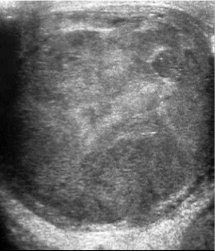
Testicular carcinoma. Ultrasound demonstrating heterogeneous echotexture throughout the testicle.
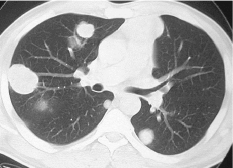
Lung metastases. CT image showing multiple lung masses in a patient with metastatic testicular seminoma.
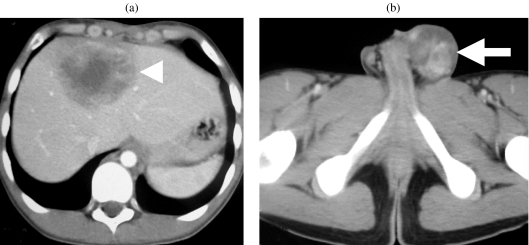
Metastatic non-seminomatous germ cell tumour. Contrast-enhanced CT shows (a) liver metastasis and (b) the left testicular tumour (arrow).
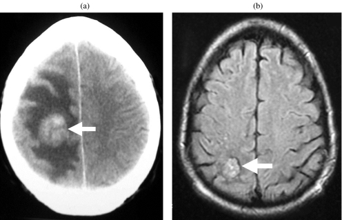
Brain metastasis. (a) Contrast-enhanced CT showing an enhancing mass (arrow) in the right parietal lobe with surrounding oedema. (b) FLAIR MR image following treatment shows the lesion to be smaller and the surrounding oedema to have resolved.
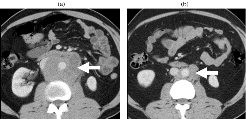
Retroperitoneal lymphadenopathy. Contrast-enhanced CT (a) before and (b) after chemotherapy shows retroperitoneal lymphadenopathy (arrow) encasing the aorta. The post-chemotherapy scan shows significant reduction in the burden of retroperitoneal lymphadenopathy.
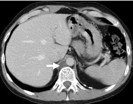
Mature teratoma differentiated in retrocrural node (arrow). Contrast-enhanced CT shows low density lesion in the right retrocrural region.
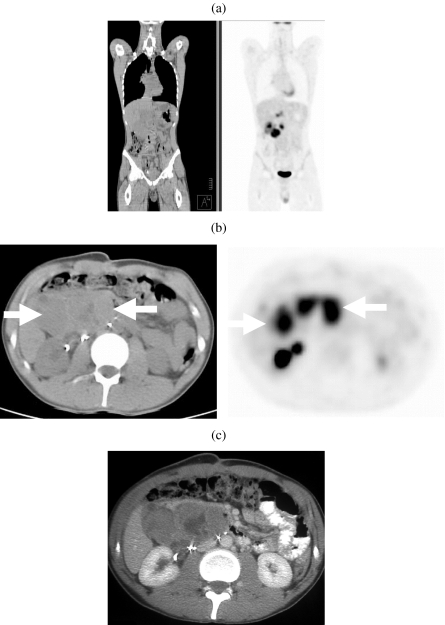
Non-seminomatous germ cell tumour. (a) Coronal and (b) axial images on 18-FDG enhanced PETCT and (c) iodinated contrast enhanced CT showing a large metabolically active retroperitoneal mass (arrows).
References
-
- Coakley FV, Hricak H, Presti JC Jr. Imaging and management of atypical testicular masses. Urol Clin North Am. 1998;25:375–88. - PubMed
-
- Guthrie JA, Fowler RC. Ultrasound diagnosis of testicular tumours presenting as epididymal disease. Clin Radiol. 1992;46:397–400. - PubMed
-
- Richie JP, Steele GS. Neoplasms of the testis. In: Walsh PC, Retik AB, Vaughan ED, editors. Campbell’s Urology. 8th edn. Philadelphia., PA: Saunders; 2001. pp. 2876–919.
-
- Grantham JG, Charboneau JW, James EM, Kirschling RJ, Kvols LK, Segura JW, et al. Testicular neoplasms: 29 tumors studied by high-resolution US. Radiology. 1985;157:775–80. - PubMed
-
- Senay BA, Stein BS. Testicular neoplasm diagnosed by ultrasound. J Surg Oncol. 1986;32:110–12. - PubMed
MeSH terms
LinkOut - more resources
Full Text Sources
Medical