. 2006 Sep;114(9):1367-73.
doi: 10.1289/ehp.8906.

Apoptosis and Bax expression are increased by coal dust in the polycyclic aromatic hydrocarbon-exposed lung

Affiliations

Apoptosis and Bax expression are increased by coal dust in the polycyclic aromatic hydrocarbon-exposed lung

Mohamed M Ghanem et al. Environ Health Perspect. 2006 Sep.

Abstract

Background: Miners inhaling respirable coal dust (CD) frequently develop coal workers' pneumoconiosis, a dust-associated pneumoconiosis characterized by lung inflammation and variable fibrosis. Many coal miners are also exposed to polycyclic aromatic hydrocarbon (PAH) components of diesel engine exhaust and cigarette smoke, which may contribute to lung disease in these workers. Recently, apoptosis was reported to play a critical role in the development of another pneumoconiosis of miners, silicosis. In addition, CD was reported to suppress cytochrome P450 1A1 (CYP1A1) induction by PAHs.

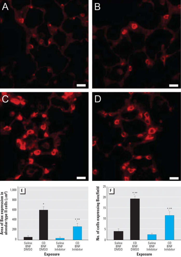
Methods: We investigated the hypothesis that apoptosis plays a critical role in lung injury and down-regulation of CYP1A1 induction in mixed exposures to CD and PAHs. We exposed rats intratracheally to 0.0, 2.5, 10.0, 20.0, or 40.0 mg/rat CD and, 11 days later, to intraperitoneal beta-naphthoflavone (BNF) , a PAH. In another group of rats exposed to CD and BNF, caspase activity was inhibited by injection of the pan-caspase inhibitor Q-VD-OPH [quinoline-Val-Asp (OMe) -CH2-OPH].

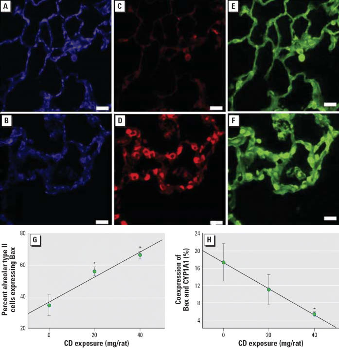
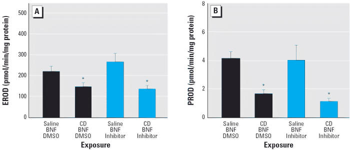
Results: In rats exposed to BNF, CD exposure increased alveolar expression of the proapoptotic mediator Bax but decreased CYP1A1 induction relative to BNF exposure alone. Pan-caspase inhibition decreased CD-associated Bax expression and apoptosis but did not restore CYP1A1 activity. Further, CD-induced lung inflammation and alveolar epithelial cell hypertrophy and hyperplasia were not suppressed by caspase inhibition.

Conclusions: Combined BNF and CD exposure increased Bax expression and apoptosis in the lung, but Bax and apoptosis were not the major determinants of early lung injury in this model.


Figures

Figure 1
Figure 1
Quantification (mean ± SE) of Bax expression in the proximal alveolar region with increasing CD exposure in BNF-exposed rats. (A) Area of Bax expression. (B) The number of cells positive for Bax. *Significantly different from saline/BNF (p ≤ 0.05).
Figure 2
Figure 2
Triple-label immunofluorescence for CYP1A1, Bax, and cytokeratin 8/18 in the BNF-exposed pulmonary alveolus in response to CD exposure. (A) CYP1A1 expression in a BNF-exposed rat. (B) CYP1A1 expression in a rat exposed to CD and BNF. (C) Bax expression in a BNF-exposed rat. (D) Increased Bax expression in a CD- and BNF-exposed rat. (E) Expression of cytokeratin 8/18 (alveolar type II cell marker) in a BNF-exposed rat. (F) Increased expression of cytokeratin 8/18 in a BNF- and CD-exposed rat. Blue fluorescence indicates CYP1A1, red indicates Bax, and green indicates cytokeratin 8/18; bars = 20 μm. (G) The percentage of alveolar type II cells expressing Bax is increased by CD exposure. (H) The percentage of alveolar type II cells expressing both Bax and CYP1A1 is decreased by CD exposure. Values shown are mean ± SE. *Significantly different from saline/BNF (p ≤ 0.05).
Figure 3
Figure 3
Immunofluorescent staining showing suppression of Bax expression by Q-VD-OPH in the proximal alveolar region of rats receiving (A) saline/BNF/DMSO, (B) saline/BNF/caspase inhibitor, (C) CD/BNF/DMSO, or (D) CD/BNF/caspase inhibitor. Bars = 20 μm. (E) Bax expression area localized to alveolar type II cells. (F) The number of Bax-expressing cells. Values shown are mean ± SE. *Significantly different from saline/BNF (p ≤ 0.05). **Significantly different from corresponding rats not receiving Q-VD-OPH.
Figure 4
Figure 4
CYP1A1 immunofluorescence (mean ± SE) in the alveolar region of BNF-induced rats exposed to CD with and without pan-caspase inhibition by Q-VD-OPH. (A) CYP1A1 expression in alveolar cells that are not type II cells. (B) Proportional CYP1A1 expression area in alveolar type II cells. *Significantly different from saline/BNF (p ≤ 0.05).
Figure 5
Figure 5
Morphologic features of caspase inhibition in CD-exposed pulmonary alveolus. (A,B) Lung photomicrographs demonstrating dust-laden macrophages in alveolar spaces (yellow arrows) and in the interstitium (blue arrows), with hypertrophy and hyperplasia of alveolar type II cells (arrowheads) from (A) a CD-exposed rat (CD/BNF/DMSO) and (B) a CD-exposed rat following caspase inhibition (CD/BNF/inhibitor). (C,D) TUNEL assay showing green fluorescent apoptotic cells (arrows) in (C) a CD-exposed rat (CD/BNF/DMSO) and (D) a CD-exposed rat after caspase inhibition (CD/BNF/inhibitor); note the decreased number of apoptotic cells compared with (C). Bars = 20 μm. (E) Semiquantitative histopathology scores with no evidence that caspase inhibition modifies CD-induced alveolitis or alveolar epithelial cell hypertrophy and hyperplasia. (F) Apoptosis in BNF-treated rats exposed to IT CD with and without caspase inhibition. Apoptosis is significantly higher in the proximal alveolar (PA) region when compared with random alveolar (RA) regions. CD significantly increased apoptosis; caspase inhibition significantly decreased, but did not entirely abrogate, CD-induced apoptosis. Values shown are mean ± SE. *Significantly different from random alveolar region (p ≤ 0.05). **Significantly different from corresponding rats not receiving CD. ***Significantly different from corresponding rats not receiving Q-VD-OPH.
Figure 6
Figure 6
CD significantly decreases both CYP1A1- and CYP2B1-dependent metabolism in the lung, shown by EROD activity (A) and PROD activity (B) of the BNF-exposed lung. Values shown are mean ± SE. *Significantly different from rats not exposed to CD (p ≤ 0.05).
Figure 7
Figure 7
Effect of CD on lung CYP1A1 Western blots. (A) Representative Western blots of lung microsomes. In the top panel, lane 1 is the CYP1A1 control, lanes 2–7 are from BNF-exposed rats, lanes 8–13 are from BNF- and CD-exposed rats, and lane 14 is the molecular weight marker. In the bottom panel, lane 1 is the CYP1A1 control, lanes 2–6 are from rats exposed to BNF and caspase inhibitor; lanes 7–12 are from rats exposed to BNF, CD, and caspase inhibitor; lane 13 is from a rat exposed to both CD and BNF, and lane 14 is the molecular weight marker. (B) Densitometry values (mean ± SE). *Significantly different from rats not exposed to CD (p ≤ 0.05).

Similar articles

Cited by

References

    1. Barazzone C, Horowitz S, Donati YR, Rodriguez I, Piguet PF. Oxygen toxicity in mouse lung: pathways to cell death. Am J Respir Cell Mol Biol. 1998;19:573–581. - PubMed
    1. Borges VM, Lopes MF, Falcao H, Leite-Junior JH, Rocco PR, Davidson WF, et al. Apoptosis underlies immuno-pathogenic mechanisms in acute silicosis. Am J Respir Cell Mol Biol. 2002;27:78–84. - PubMed
    1. Burke MD, Thompson S, Elcombe CR, Halpert J, Haaparanta T, Mayer RT. Ethoxy-, pentoxy-, and benzyloxyphenoxazones and homologues: a series of substrates to distinguish between different induced cytochromes P-450. J Biochem Pharmacol. 1985;34:3337–3345. - PubMed
    1. Caserta TM, Smith AN, Gultice AD, Reedy MA, Brown TL. Q-VD-OPh, a broad spectrum caspase inhibitor with potent antiapoptotic properties. Apoptosis. 2003;8:345–352. - PubMed
    1. Douglas AN, Robertson A, Chapman JS, Ruckley VA. Dust exposure, dust recovered from the lung, and associated pathology in a group of British coalminers. Br J Ind Med. 1986;43:795–801. - PMC - PubMed

Publication types

MeSH terms

LinkOut - more resources