

Exit from arsenite-induced mitotic arrest is p53 dependent
- PMID: 16966095
- PMCID: PMC1570045
- DOI: 10.1289/ehp.8969
Exit from arsenite-induced mitotic arrest is p53 dependent
Abstract
Background: Arsenic is both a human carcinogen and a chemotherapeutic agent, but the mechanism of neither arsenic-induced carcinogenesis nor tumor selective cytotoxicity is clear. Using a model cell line in which p53 expression is regulated exogenously in a tetracycline-off system (TR9-7 cells) , our laboratory has shown that arsenite disrupts mitosis and that p53-deficient cells [p53(-)], in contrast to p53-expressing cells [p53(+)], display greater sensitivity to arsenite-induced mitotic arrest and apoptosis.
Objective: Our goal was to examine the role p53 plays in protecting cells from arsenite-induced mitotic arrest.
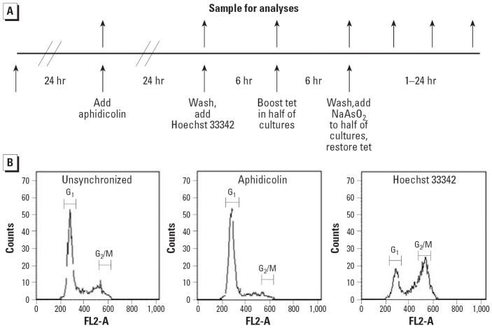
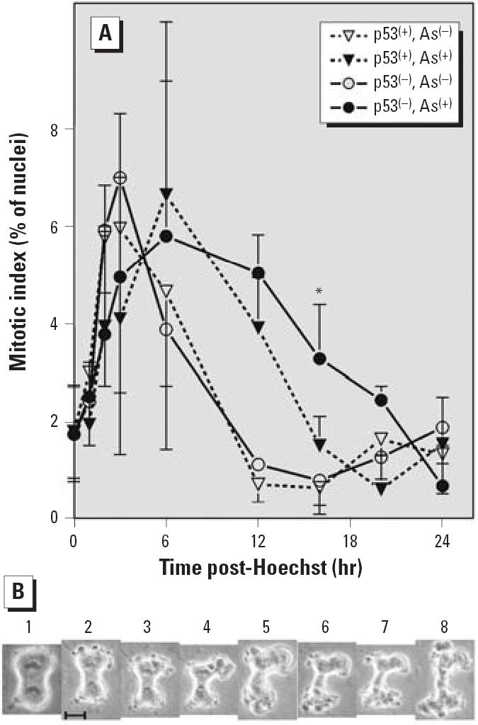
Methods: p53(+) and p53(-) cells were synchronized in G2 phase using Hoechst 33342 and released from synchrony in the presence or absence of 5 microM sodium arsenite.
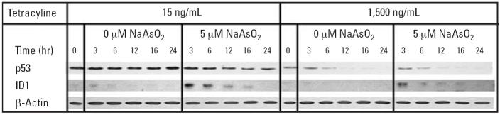
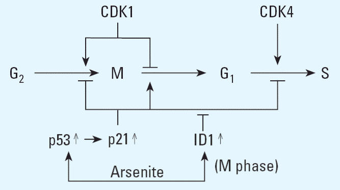
Results: Mitotic index analysis demonstrated that arsenite treatment delayed exit from G2 in p53(+) and p53(-) cells. Arsenite-treated p53(+) cells exited mitosis normally, whereas p53(-) cells exited mitosis with delayed kinetics. Microarray analysis performed on mRNAs of cells exposed to arsenite for 0 and 3 hr after release from G2 phase synchrony showed that arsenite induced inhibitor of DNA binding-1 (ID1) differentially in p53(+) and p53(-) cells. Immunoblotting confirmed that ID1 induction was more extensive and sustained in p53(+) cells.
Conclusions: p53 promotes mitotic exit and leads to more extensive ID1 induction by arsenite. ID1 is a dominant negative inhibitor of transcription that represses cell cycle regulatory genes and is elevated in many tumors. ID1 may play a role in the survival of arsenite-treated p53(+) cells and contribute to arsenic carcinogenicity.
Figures
Similar articles
-
p53 suppression of arsenite-induced mitotic catastrophe is mediated by p21CIP1/WAF1.J Pharmacol Exp Ther. 2006 Jul;318(1):142-51. doi: 10.1124/jpet.106.103077. Epub 2006 Apr 13. J Pharmacol Exp Ther. 2006. PMID: 16614167
-
ATM/ATR-related checkpoint signals mediate arsenite-induced G2/M arrest in primary aortic endothelial cells.Arch Toxicol. 2006 Dec;80(12):804-10. doi: 10.1007/s00204-006-0110-4. Arch Toxicol. 2006. PMID: 16645841
-
Depletion of securin increases arsenite-induced chromosome instability and apoptosis via a p53-independent pathway.Toxicol Sci. 2006 Mar;90(1):73-86. doi: 10.1093/toxsci/kfj070. Epub 2005 Dec 7. Toxicol Sci. 2006. PMID: 16338954
-
[Cell cycle regulation after exposure to ionizing radiation].Bull Cancer. 1999 Apr;86(4):345-57. Bull Cancer. 1999. PMID: 10341340 Review. French.
-
Disruption of Mitotic Progression by Arsenic.Biol Trace Elem Res. 2015 Jul;166(1):34-40. doi: 10.1007/s12011-015-0306-7. Epub 2015 Mar 22. Biol Trace Elem Res. 2015. PMID: 25796515 Free PMC article. Review.
Cited by
-
The NRF2-mediated oxidative stress response pathway is associated with tumor cell resistance to arsenic trioxide across the NCI-60 panel.BMC Med Genomics. 2010 Aug 13;3:37. doi: 10.1186/1755-8794-3-37. BMC Med Genomics. 2010. PMID: 20707922 Free PMC article.
-
Overexpression of hsa-miR-186 induces chromosomal instability in arsenic-exposed human keratinocytes.Toxicol Appl Pharmacol. 2019 Sep 1;378:114614. doi: 10.1016/j.taap.2019.114614. Epub 2019 Jun 6. Toxicol Appl Pharmacol. 2019. PMID: 31176655 Free PMC article.
-
Integration of microRNAome, proteomics and metabolomics to analyze arsenic-induced malignant cell transformation.Oncotarget. 2017 Jun 27;8(53):90879-90896. doi: 10.18632/oncotarget.18741. eCollection 2017 Oct 31. Oncotarget. 2017. PMID: 29207610 Free PMC article.
-
Subhepatotoxic exposure to arsenic enhances lipopolysaccharide-induced liver injury in mice.Toxicol Appl Pharmacol. 2008 Jan 15;226(2):128-39. doi: 10.1016/j.taap.2007.08.020. Epub 2007 Aug 31. Toxicol Appl Pharmacol. 2008. PMID: 17919673 Free PMC article.
-
Arsenite-induced mitotic death involves stress response and is independent of tubulin polymerization.Toxicol Appl Pharmacol. 2008 Jul 15;230(2):235-46. doi: 10.1016/j.taap.2008.02.030. Epub 2008 Mar 14. Toxicol Appl Pharmacol. 2008. PMID: 18485433 Free PMC article.
References
-
- Abraham NG. Therapeutic applications of human heme oxygenase gene transfer and gene therapy. Curr Pharm Des. 2003;9:2513–2524. - PubMed
Publication types
MeSH terms
Substances
Grants and funding
LinkOut - more resources
Full Text Sources
Other Literature Sources
Molecular Biology Databases
Research Materials
Miscellaneous
