. 2006 Nov;74(11):6339-47.
doi: 10.1128/IAI.00982-06. Epub 2006 Sep 11.

Human monoclonal antibodies directed against toxins A and B prevent Clostridium difficile-induced mortality in hamsters

Affiliations

Human monoclonal antibodies directed against toxins A and B prevent Clostridium difficile-induced mortality in hamsters

Gregory J Babcock et al. Infect Immun. 2006 Nov.

Abstract

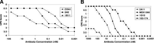
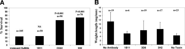
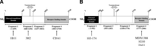
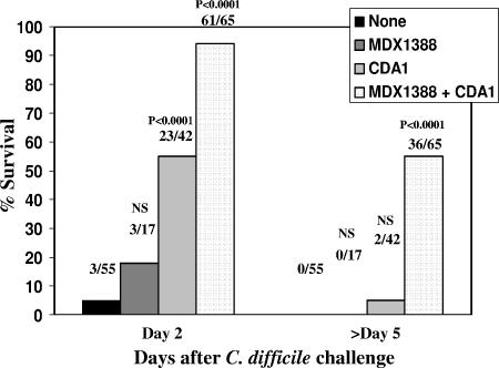
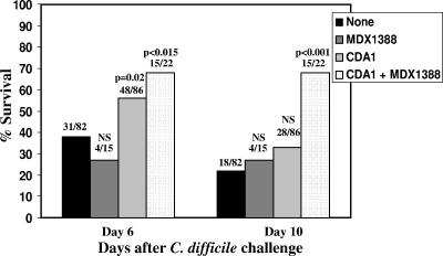
Clostridium difficile is the leading cause of nosocomial antibiotic-associated diarrhea, and recent outbreaks of strains with increased virulence underscore the importance of identifying novel approaches to treat and prevent relapse of Clostridium difficile-associated diarrhea (CDAD). CDAD pathology is induced by two exotoxins, toxin A and toxin B, which have been shown to be cytotoxic and, in the case of toxin A, enterotoxic. In this report we describe fully human monoclonal antibodies (HuMAbs) that neutralize these toxins and prevent disease in hamsters. Transgenic mice carrying human immunoglobulin genes were used to isolate HuMAbs that neutralize the cytotoxic effects of either toxin A or toxin B in cell-based in vitro neutralization assays. Three anti-toxin A HuMAbs (3H2, CDA1, and 1B11) could all inhibit the enterotoxicity of toxin A in mouse intestinal loops and the in vivo toxicity in a systemic mouse model. Four anti-toxin B HuMAbs (MDX-1388, 103-174, 1G10, and 2A11) could neutralize cytotoxicity in vitro, although systemic toxicity in the mouse could not be neutralized. Anti-toxin A HuMAb CDA1 and anti-toxin B HuMAb MDX-1388 were tested in the well-established hamster model of C. difficile disease. CDA1 alone resulted in a statistically significant reduction of mortality in hamsters; however, the combination treatment offered enhanced protection. Compared to controls, combination therapy reduced mortality from 100% to 45% (P<0.0001) in the primary disease hamster model and from 78% to 32% (P<0.0001) in the less stringent relapse model.


Figures

FIG. 1.
FIG. 1.
In vitro neutralization of cytotoxicity. (A) C. difficile toxin A (6 ng/well) was mixed with either HuMAb CDA1, 3H2, or 1B11 at various antibody concentrations from 100 to 0.001 nM. IMR-90 cells were plated at 1 × 10⁵ cells/well in a 96-well microtiter plate, and toxin A-antibody mixtures were applied to the cells. IMR-90 cells with a toxin A-antibody mixture applied were incubated for 24 h and scored for cytopathic effect (CPE) visually on a scale of 0 to 4, where 0 represents no observed toxicity and 4 indicates that 100% of the monolayer was effected by toxin A. (B) The experiment was performed as described for panel A, with the exception that C. difficile toxin B (20 pg/well) was mixed with either HuMAb 2A11, MDX1388, 1G10, or 103-174 at various antibody concentrations from 1,000 to 0.0001 nM prior to being applied to IMR-90 cells.
FIG. 2.
FIG. 2.
Neutralization of in vivo toxicity in mice. (A) Mice were treated with 100 to 250 μg either of an irrelevant HuMAb or of 1B11, CDA1, or 3H2 24 h prior to intraperitoneal administration of 100 ng of toxin A. Animals were observed for up to 72 h, and the percentages of surviving animals were plotted. The number of animals tested in each group is shown above the respective column in the bar graph, with P values indicated (the Fisher exact test). NS, not significant. (B) Mice were injected i.p. with 500 μg to 2 mg of 1B11, CDA1, or 3H2 24 h prior to ileal loop surgery. As an untreated positive control, mice were injected i.p. with PBS rather than antibody (no antibody). As a negative control for fluid accumulation due to surgery, PBS was injected into the ligated loop in place of toxin A (no toxin). The number of animals in each group is shown above the respective column in the bar graph. Toxin A (0.45 μg) was injected into ligated ileal loops of treated animals, and loops were excised 4 hours later. Weight-to-length ratios were calculated and plotted.
FIG. 3.
FIG. 3.
Domain recognition of toxin A- and toxin B-reactive HuMAbs. (A) Schematic representation of recombinant fragments cloned and expressed in E. coli, with amino acid numbers listed above and below the figure. Specific antibodies are listed below the recognized domain. (B) Schematic of recombinant toxin B fragments that were cloned and expressed in E. coli, with amino acid numbers listed above and below the figure. Specific antibodies are listed below the recognized domain.
FIG. 4.
FIG. 4.
Protection of hamsters with HuMAbs in the primary challenge model. Hamsters were treated with CDA1 (50 mg/kg/day) alone or in combination with MDX1388 (10 to 50 mg/kg/day) for 4 days beginning 3 days prior to spore administration. Clindamycin (10 mg/kg) was injected intraperitoneally 24 h prior to spore challenge. Spores were delivered orogastrically, and animals were observed for up to 11 days. A total of six experiments were performed, and a summary of all hamster primary challenge experiments using percent survival at either 2 or >5 days after challenge is shown. The fractions above the bar graphs represent the numbers of surviving animals (numerators) and the total numbers of animals (denominators), with P values indicated (the Fisher exact test). NS, not significant.
FIG. 5.
FIG. 5.
Protection of hamsters with HuMAbs in the relapse model. Clindamycin (10 mg/kg) was injected intraperitoneally 24 h prior to spore challenge. Spores were administered orogastrically, at which time vancomycin treatment (10 mg/kg/day) was initiated and continued for a subsequent 2 days. Hamsters were treated with CDA1 (60 mg/kg/day) in combination with MDX1388 (20 mg/kg/day) for 4 days beginning 2 days following spore administration. Animals were observed for mortality until day 10. A summary of all hamster relapse model experiments using percent survival at either 6 or 10 days following challenge was graphed. The fractions above the bar graphs represent the numbers of surviving animals (numerators) and the total numbers of animals (denominators), with P values indicated (the Fisher exact test). NS, not significant.

References

    1. Bartlett, J. G. 1981. Antimicrobial agents implicated in Clostridium difficile toxin-associated diarrhea of colitis. Johns Hopkins Med. J. 149:6-9. - PubMed
    1. Bartlett, J. G. 2002. Clinical practice. Antibiotic-associated diarrhea. N. Engl. J. Med. 346:334-339. - PubMed
    1. Bartlett, J. G., T. Chang, N. S. Taylor, and A. B. Onderdonk. 1979. Colitis induced by Clostridium difficile. Rev. Infect. Dis. 1:370-378. - PubMed
    1. Bartlett, J. G., T. W. Chang, M. Gurwith, S. L. Gorbach, and A. B. Onderdonk. 1978. Antibiotic-associated pseudomembranous colitis due to toxin-producing clostridia. N. Engl. J. Med. 298:531-534. - PubMed
    1. Bartlett, J. G., A. B. Onderdonk, R. L. Cisneros, and D. L. Kasper. 1977. Clindamycin-associated colitis due to a toxin-producing species of Clostridium in hamsters. J. Infect. Dis. 136:701-705. - PubMed

MeSH terms

LinkOut - more resources