. 2006 Sep 11;174(6):827-38.
doi: 10.1083/jcb.200512066.

PKA-activated ApAF-ApC/EBP heterodimer is a key downstream effector of ApCREB and is necessary and sufficient for the consolidation of long-term facilitation

Affiliations

PKA-activated ApAF-ApC/EBP heterodimer is a key downstream effector of ApCREB and is necessary and sufficient for the consolidation of long-term facilitation

Jin-A Lee et al. J Cell Biol. .

Abstract

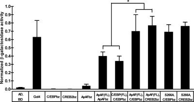
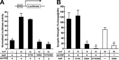
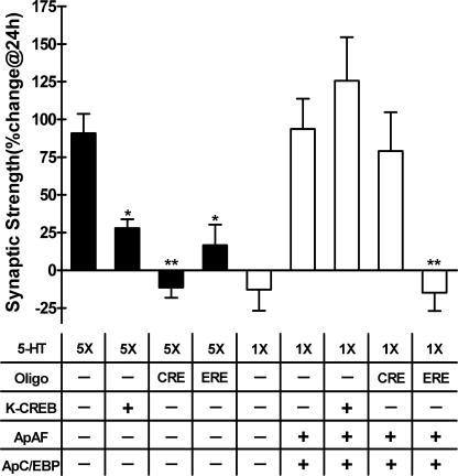
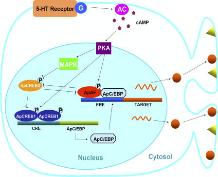
Long-term memory requires transcriptional regulation by a combination of positive and negative transcription factors. Aplysia activating factor (ApAF) is known to be a positive transcription factor that forms heterodimers with ApC/EBP and ApCREB2. How these heterodimers are regulated and how they participate in the consolidation of long-term facilitation (LTF) has not, however, been characterized. We found that the functional activation of ApAF required phosphorylation of ApAF by PKA on Ser-266. In addition, ApAF lowered the threshold of LTF by forming a heterodimer with ApCREB2. Moreover, once activated by PKA, the ApAF-ApC/EBP heterodimer transactivates enhancer response element-containing genes and can induce LTF in the absence of CRE- and CREB-mediated gene expression. Collectively, these results suggest that PKA-activated ApAF-ApC/EBP heterodimer is a core downstream effector of ApCREB in the consolidation of LTF.


Figures

Figure 1.
Figure 1.
Interaction between the nuclear protein ApAF and ApC/EBP or ApCREB2 in an A. kurodai neuron. The histogram represents the degree of interaction in terms of normalized β-galactosidase activity (β-galactosidase/luciferase activity). ApAF or ApC/EBP can form homodimers through bZIP (n = 9 and n = 6, respectively). ApAF can also form heterodimers with ApC/EBP (n = 4) or with ApCREB2 (n = 4). Heterodimerization seems to be more favorable than homodimerization (*, P < 0.05; one-way analysis of variance (ANOVA) and Newman-Keuls multiple comparison test for ApAF(FL)-ApAFbz, C/EBP(FL)-C/EBPbz, ApAF(FL)-C/EBPbz, and ApAF(FL)-CREB2bz). ApAF (S266A) has a similar binding affinity with ApC/EBP (n = 3) and ApCREB2 (n = 3) as ApAF(WT). In contrast, neither the pair alone (n = 6) nor the Gal4DB-bait (bZIP of ApC/EBP [n = 6], ApCREB2 [n = 7], or ApAF [n = 3]) on its own could activate reporter genes. Gal4 is used as a positive control (n = 5). The bars correspond to normalized mean β-galactosidase activity ± the SEM. BD and AD represent the DNA-binding domain and transcription activation domain, respectively. bz represents the bZIP domain of the transcription factors. AD, BD, ApAF (FL), C/EBP (FL), C/EBPbz, CREB2bz, ApAFbz, and S266A represent Gal4AD, Gal4BD, Gal4AD-ApAF (full-length), Gal4AD-ApC/EBP (full-length), Gal4BD-ApC/EBPbz, Gal4BD-ApCREB2bz, Gal4BD-ApAFbz, and ApAF (S266A), respectively.
Figure 2.
Figure 2.
ApAF induced ERE-mediated gene expression and LTF by interacting with ApC/EBP. (A) Transcriptional activity of ApAF, ApC/EBP, or ApAF–ApC/EBP via the ERE sequence in A. kurodai sensory neurons. +, DNA constructs injected with ERE-luciferase reporter into sensory cells in the pleural ganglia. Either one (1×) or five pulses (5×) of 5-HT were applied to sensory cells. ERE-driven reporter gene expression was increased by ApAF (n = 4), ApC/EBP (n = 4), or ApAF–ApC/EBP (n = 4) overexpression in comparison to nonexpressing control cells (n = 4; ***, P < 0.001; one-way ANOVA and Newman-Keuls multiple comparison test for groups indicated by open bars). The reporter gene expression was further enhanced by 5-HT treatment in ApAF- (n = 4), ApC/EBP- (n = 5), and ApAF–ApC/EBP-introduced (n = 8) cells (*, P < 0.05; **, P < 0.01; ***, P < 0.001; two-tailed unpaired t test). Each bar corresponds to normalized mean luciferase activity ± the SEM. Five pulses of 5-HT treatment significantly increased ERE-mediated gene expression (n = 4), whereas one pulse of 5-HT treatment did not (n = 5). (B) The effect and specificity of ApAF dsRNA. The expression level of ApAF mRNA was examined by in situ hybridization. The expression level of ApAF was significantly lower in ApAF dsRNA–injected neurons (n = 5) than in luciferase dsRNA–injected neurons (n = 9; *, P < 0.05; two-tailed unpaired t test), whereas the expression level of ApCREB1a was not affected by ApAF dsRNA injection (n = 6). The percentage of change in mRNA is calculated as the relative expression level of ApAF or ApCREB1a mRNA in ApAF dsRNA–injected neurons compared with luciferase dsRNA–injected neurons. The bar represents the mean ± the SEM. Bar, 25 μm. (C) The effect of ApAF–ApC/EBP heterodimer on LTF. +, DNA constructs injected into sensory cells. EPSPs were measured before and 24 h after one or five pulses of 5-HT were applied to the sensory-to-motor synapse. For shaded bars, injection of ApAF dsRNA completely blocked both the LTF induced by five pulses of 5-HT (n = 11) and the LTF induced by ApC/EBP overexpression combined with one pulse of 5-HT (n = 8), whereas the injection of luciferase dsRNA did not block either type of LTF (5×5-HT, n = 8; 1×5-HT, n = 4; **, P <0.01; one way ANOVA and Newman-Keuls multiple comparison test). For open bars, ApAF overexpression combined with one pulse of 5-HT did not produce LTF (n = 8), whereas ApC/EBP overexpression combined with 5-HT did (n = 13). However, ApAF overexpression enhanced the LTF induced by ApC/EBP and one pulse of 5-HT (n = 17; *, P < 0.05). The noninjected control cells produced normal LTF by five pulses of 5-HT (n = 10). Representative examples of recording traces are shown on the right. (D) STF was not affected by ApAF dsRNA microinjection. The mean percentage of changes in EPSP of ApAF dsRNA–injected cells (n = 4) was not significantly different from that of noninjected control cells (n = 4) or luciferase dsRNA–injected cells (n = 4; P > 0.05; one way ANOVA and Newman-Keuls multiple comparison test). Bars correspond to mean percentage changes ± the SEM in EPSP amplitudes.
Figure 3.
Figure 3.
ApAF phosphorylation by PKA is involved in ApAF-mediated LTF. (A) The effect of PKA activation on ApAF-mediated LTF. +, DNA constructs injected into sensory cells of sensory-to-motor synapses. The cultures were exposed to one (1×) or five pulses (5×) of 5-HT. Incubation with 10 μM KT5720 for 2 h completely blocked the LTF induced by one pulse of 5-HT with ApC/EBP overexpression (n = 4) or with overexpression of ApAF and ApC/EBP (n = 4), as well as the LTF induced by five pulses of 5-HT (n = 5; *, P < 0.05; **, P < 0.01; ***, P < 0.001; two-tailed Mann-Whitney test). Bars correspond to mean percentage changes ± the SEM in EPSP amplitudes. The data without inhibitor treatment are the same as in Fig. 2 C. (B) Phosphorylation of ApAF. (top) Purified ApAF was incubated with Aplysia cell lysate as a source of kinases. PKA inhibitor KT5720 dramatically blocked the phosphorylation of ApAF, whereas the MEK inhibitor PD98059 or the PKC inhibitor chelerythrine did not. (bottom) ApAF proteins were purified from E. coli as GST fusion proteins and were incubated with catalytic subunits of PKA. WT and S175A mutant were directly phosphorylated by PKA, whereas S266A mutant and S175/266A double mutant were not. This phosphorylation was completely blocked by KT5720.
Figure 4.
Figure 4.
ApAF phosphorylation by PKA on Ser-266 is essential for ERE-mediated gene expression and LTF. (A) The effect of ApAF phosphorylation by PKA at Ser-266 on ERE-mediated gene expression. +, DNA constructs injected with ERE-luciferase into sensory cells in the pleural ganglia. 24 h after microinjection, the cells were treated with one pulse (1×) of 5-HT, which does not induce gene expression. ERE-driven reporter gene expressions by the introduction of ApAF(S266A) and ApAF(S175/266A) were completely blocked (both n = 4; *, P < 0.001; one-way ANOVA and Newman-Keuls multiple comparison test). Each bar corresponds to the mean normalized luciferase activity ± the SEM. The experimental data for WT ApAF are the same as in Fig. 2 A. (B) The effect of ApAF phosphorylation by PKA at Ser-266 on LTF. +, DNA constructs injected into the sensory cell of sensory-to-motor synapses. The cultures were exposed to one (1×) or five pulses (5×) of 5-HT. Overexpression of ApAF mutants (S266A and S175/266A) blocked ApAF–ApC/EBP–mediated LTF (n = 5 and n = 8, respectively), whereas overexpression of ApAF(S175A) did not block LTF (n = 9; *, P < 0.05, **, P < 0.01; one-way ANOVA and Newman-Keuls multiple comparison test for one-pulse 5-HT groups indicated by shaded bars). Moreover, ApAF(S266A) impaired the LTF induced by five pulses of 5-HT (n = 5; **, P < 0.01; two-tailed unpaired t test for five-pulse 5-HT groups indicated by open bar). Bars correspond to mean percentage changes ± the SEM in EPSP amplitudes. The data for WT ApAF and the data without injection are the same as in Fig. 2 C.
Figure 5.
Figure 5.
ApAF phosphorylation by PKA on Ser-266 is not required to relieve repression by ApCREB2. (A) The effect of ApAF phosphorylation by PKA at Ser-266 on ApCREB2-mediated repression. +, DNA constructs injected with CRE-luciferase reporter into cells in the abdominal ganglia. ApCREB2 overexpression (n = 6) blocked CRE-driven reporter gene expression (n = 5) activated by ApCREB1. ApAF(S175A) (n = 3), ApAF(S266A) (n = 3), and ApAF(S175/266A) (n = 4), as well as ApAF WT (n = 3) restored CRE-driven gene expression repressed by ApCREB2 (*, P < 0.001; one-way ANOVA and Newman-Keuls multiple comparison test). Luciferase activity was normalized to β-galactosidase activity. Each bar corresponds to normalized mean luciferase activity ± the SEM. CRE-luciferase alone (n = 5); CRE-luciferase with ApAF (n = 8); CRE-luciferase with ApAF and ApCREB2 (n = 4). (B) The effect of ApAF phosphorylation by PKA at Ser-266 on the blockage of LTF by ApCREB2 repressor. +, DNA constructs injected into sensory cells of sensory-to-motor synapse. The cultures were treated with five pulses of 5-HT. ApCREB2 overexpression significantly impaired the LTF induced by five pulses of 5-HT (n = 12). ApAF overexpression combined with five pulses of 5-HT relieved the blockage of LTF by ApCREB2 (n = 13), whereas ApAF overexpression alone did not affect LTF (n = 9; *, P < 0.05; **, P < 0.01; one-way ANOVA and Newman-Keuls multiple comparison test for the groups represented by a hatched bar and shaded bars). Moreover, ApAF mutants (S175A [n = 5], S266A [n = 4], and S175/266A [n = 4]) also relieved the blockage of LTF by ApCREB2 (**, P < 0.01; one-way ANOVA and Newman-Keuls multiple comparison test for the groups represented by a hatched bar and open bars). For five pulses of 5-HT, control is n = 17. For ApAF and ApCREB2 overexpression without the 5-HT treatment n = 5. Bars correspond to mean percentage changes ± the SEM in EPSP amplitudes.
Figure 6.
Figure 6.
LTF produced by PKA-activated ApAF and ApC/EBP was not blocked by inhibiting CRE- and CREB-mediated gene expression, but was still blocked by inhibition of ERE-mediated gene expression. +, DNA constructs or oligonucleotides (CRE and ERE) injected into sensory cells. The cultures were exposed to one (1×) or five pulses (5×) of 5-HT. Injection of CRE oligonucleotides (oligo; n = 7), as well as K-CREB (n = 6), did not block the LTF (n = 13) produced by ApAF and ApC/EBP in the presence of one-pulse 5-HT treatment. However, injection of CRE (n = 4) or pNEXδ–K-CREB (n = 8) blocked the LTF produced by five pulses (5×) of 5-HT (*, P < 0.05; **, P < 0.01; one-way ANOVA and Newman-Keuls multiple comparison test). In contrast, injection of ERE oligonucleotides completely blocked the LTF induced by ApAF–ApC/EBP in the presence of one-pulse 5-HT (n = 6; **, P < 0.01). ERE oligonucleotides also blocked the LTF induced by five pulses of 5-HT (n = 7; *, P < 0.05). Bars correspond to mean percentage changes ± the SEM in EPSP amplitudes. 5-HT controls were 1×, n = 7; 5×, n = 13.
Figure 7.
Figure 7.
The transcriptional regulation by ApAF in consolidation of LTF. In basal state without stimulation by 5-HT, ApCREB2 represses CRE-mediated gene expression by ApCREB1. ApAF binds to ApCREB2, allowing ApCREB1 to form a homodimer, which can bind to the CRE element. Thus, ApAF can lower the threshold of LTF, producing a “primed” state for LTF. The multiple pulses of 5-HT up-regulate cAMP levels within sensory cells via G-protein–coupled receptors, and activate PKA, which then recruits another kinase, MAPK. Both PKA and MAPK translocate to the nucleus, where they phosphorylate the transcriptional factors ApCREB1/2 and ApAF. Phosphorylated ApCREB2 may dissociate easily from ApCREB1 and ApAF. Activation of ApCREB1 by PKA induces an immediate-early gene such as ApC/EBP, and then phosphorylated ApAF can form a heterodimer with newly synthesized ApC/EBP. Functional cooperation of PKA-activated ApAF–ApC/EBP heterodimer is essential for gene induction involved in the consolidation of LTF. Therefore, ApAF may serve as a powerful cofactor during consolidation of LTF.

References

    1. Abel, T., and E. Kandel. 1998. Positive and negative regulatory mechanisms that mediate long-term memory storage. Brain Res. Brain Res. Rev. 26:360–378. - PubMed
    1. Alberini, C.M., M. Ghirardi, R. Metz, and E.R. Kandel. 1994. C/EBP is an immediate-early gene required for the consolidation of long-term facilitation in Aplysia. Cell. 76:1099–1114. - PubMed
    1. Barondes, S.H., and H.D. Cohen. 1968. Memory impairment after subcutaneous injection of acetoxycycloheximide. Science. 160:556–557. - PubMed
    1. Bartsch, D., M. Ghirardi, P.A. Skehel, K.A. Karl, S.P. Herder, M. Chen, C.H. Bailey, and E.R. Kandel. 1995. Aplysia CREB2 represses long-term facilitation: relief of repression converts transient facilitation into long-term functional and structural change. Cell. 83:979–992. - PubMed
    1. Bartsch, D., A. Casadio, K.A. Karl, P. Serodio, and E.R. Kandel. 1998. CREB1 encodes a nuclear activator, a repressor, and a cytoplasmic modulator that form a regulatory unit critical for long-term facilitation. Cell. 95:211–223. - PubMed

Publication types

MeSH terms

Substances