. 2006 Sep 11;174(6):877-88.
doi: 10.1083/jcb.200603111.

RLIP76 (RalBP1) is an R-Ras effector that mediates adhesion-dependent Rac activation and cell migration

Affiliations

RLIP76 (RalBP1) is an R-Ras effector that mediates adhesion-dependent Rac activation and cell migration

Lawrence E Goldfinger et al. J Cell Biol. .

Abstract

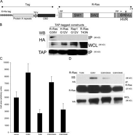
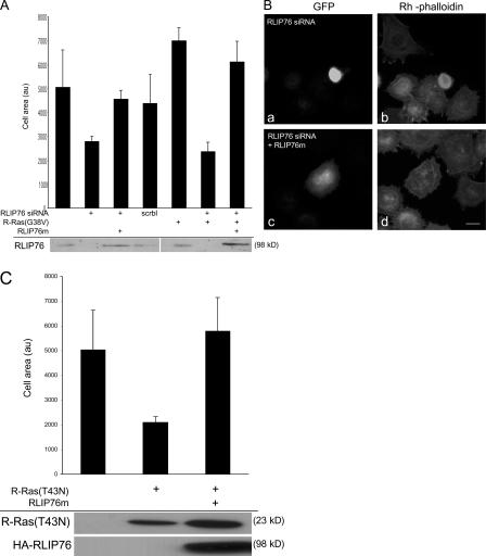
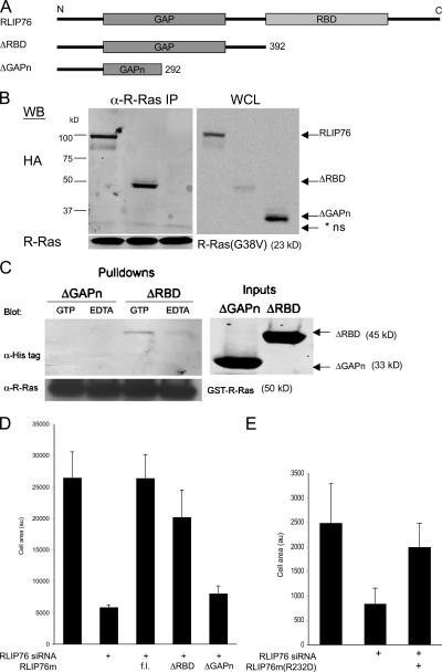
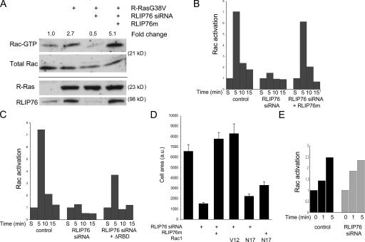
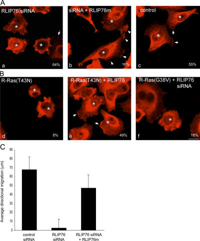
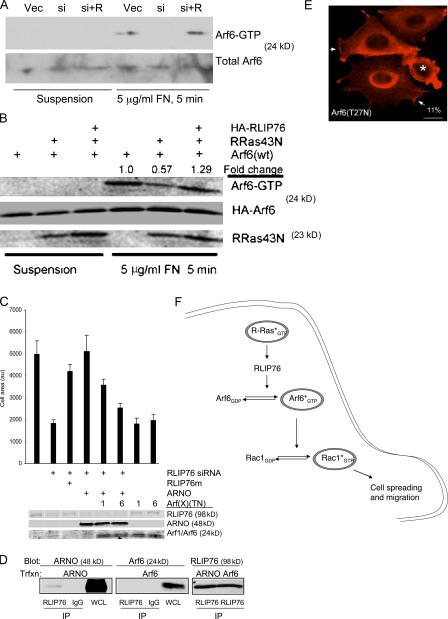
The Ras family of small GTPases regulates cell proliferation, spreading, migration and apoptosis, and malignant transformation by binding to several protein effectors. One such GTPase, R-Ras, plays distinct roles in each of these processes, but to date, identified R-Ras effectors were shared with other Ras family members (e.g., H-Ras). We utilized a new database of Ras-interacting proteins to identify RLIP76 (RalBP1) as a novel R-Ras effector. RLIP76 binds directly to R-Ras in a GTP-dependent manner, but does not physically associate with the closely related paralogues H-Ras and Rap1A. RLIP76 is required for adhesion-induced Rac activation and the resulting cell spreading and migration, as well as for the ability of R-Ras to enhance these functions. RLIP76 regulates Rac activity through the adhesion-induced activation of Arf6 GTPase and activation of Arf6 bypasses the requirement for RLIP76 in Rac activation and cell spreading. Thus, we identify a novel R-Ras effector, RLIP76, which links R-Ras to adhesion-induced Rac activation through a GTPase cascade that mediates cell spreading and migration.


Figures

Figure 1.
Figure 1.
Identification of RLIP76 as an R-Ras effector in cell spreading. (A) Schematic representation of the TAP–R-Ras fusion construct. The modified TAP tag consists of an N-terminal poly-histidine tag (6-His tag), a protein A module, the recognition sequence for the TEV protease, and a calmodulin-binding domain (CBD). The following important features of the R-Ras moiety are shown: two proline-rich SH3-binding sequences (PP), one of which is contained within the C-terminal hypervariable region (HVR); the switch 1 effector-binding loop region (SW1), which is important for effector binding; the switch 2 region (SW2), which is involved in binding guanine nucleotide; the Cys213 palmitoylation site (*) and the Cys215 methylation and geranylgeranylation site (¥); and the R-Ras isoprenylation motif. (B) TAP fusions of constitutively active variants of R-Ras, H-Ras, Rap1A, and dominant-negative R-Ras(T43N) were cotransfected with HA-RLIP76 cDNA into NIH 3T3 cells. TAP-Ras proteins were isolated from cell lysates by IgG–Sepharose affinity chromatography, and protein complexes were fractionated by SDS-PAGE and Western blotted with antibodies to the TAP tag (α-MLCKsk) or HA. HA-RLIP76 was detected in R-Ras(G38V) precipitates, but not in precipitates of H-Ras(G12V), Rap1A(G12V), or R-Ras(T43N), indicating that RLIP76 binds in a GTP-dependent manner exclusively to R-Ras among the members of this Ras subfamily. IP, immunoprecipitate; WCL, whole cell lysate. (C) NIH 3T3 cells were cotransfected with cDNA encoding GFP and the following variants of human R-Ras: constitutively active (G38V), dominant-negative (T43N), and effector loop mutants (D64A and D64E) on the activated G38V background. Cells were plated onto fibronectin-coated coverslips, fixed after 45 min, and stained with rhodamine-phalloidin to reveal F-actin. The cell periphery, as delineated by phalloidin labeling, was used to calculate the area of GFP-positive cells. At least 40 cells were counted for each sample. Mean area measurements are representative of at least three independent experiments and are shown ± the SEM. (D) HA-RLIP76 was cotransfected with the indicated R-Ras protein variants in NIH 3T3 cells. R-Ras was immunoprecipitated from each sample with anti–R-Ras antibodies, and the bound proteins were eluted in SDS buffer and separated by SDS-PAGE, and then Western blotted with antibodies to R-Ras or to HA. HA-RLIP76 was detected in R-Ras(G38V) and -G38VD64A precipitates, but not in -T43N or -G38VD64E precipitates.
Figure 2.
Figure 2.
RLIP76 is required for R-Ras–induced cell spreading. (A) NIH 3T3 cells were transfected with R-Ras(G38V) in the presence or absence of 50 pmol of a double-stranded siRNA targeting both human and mouse RLIP76 (RLIP76 siRNA; Rosse et al., 2003) or a sequence-scrambled siRNA (scrbl). Cell lysates were blotted with anti-RLIP76 antibodies. RLIP76 expression was restored in siRNA-transfected cells by coexpression of RLIP76 cDNA containing three silent mutations in the siRNA target site (RLIP76m). Cells were plated on fibronectin-coated coverslips and cell area was measured as described in Materials and methods. Mean area measurements of GFP-positive cells are representative of at least three independent experiments and are shown ± the SEM. (B) Representative micrographs (a and c, GFP to mark transfected cells; b and d, rhodamine-phalloidin to detect F-actin) of cells spreading on fibronectin; cells were transfected with RLIP76 siRNA in the absence (a and b) or presence of the RLIP76m rescue construct (c and d). Bar, 5 μm. (C) NIH 3T3 cells were transfected with dominant-negative R-Ras(T43N), with or without cotransfection with RLIP76, and cell spreading was measured as described in Materials and methods. Mean area measurements of GFP-positive cells are representative of at least three independent experiments and are shown ± the SEM. Expression levels of the transfected proteins were assayed by Western blotting with antibodies to R-Ras (top) or HA (bottom).
Figure 3.
Figure 3.
The RhoGAP, but not the RBD of RLIP76, is required for binding to R-Ras and for cell spreading. (A) Schematic diagram of RLIP76 truncation mutants. Each of the indicated constructs was generated with N-terminal HA tags for mammalian expression and N-terminal His tags for bacterial expression and purification. (B) Coimmunoprecipitation of RLIP76 truncation mutants and R-Ras. Full-length RLIP76 and RLIP76ΔRBD coprecipitated with activated R-Ras, but the RLIP76ΔGAPn truncation did not. IP, immunoprecipitates; WCL, whole cell lysate; * ns, nonspecific. (C) In vitro binding of RLIP76ΔRBD to activated R-Ras. GST–R-Ras was coupled to GSH–Sepharose and activated by addition of excess GTP or was kept nucleotide-free in 10 mM EDTA before addition of purified RLIP76 proteins. RLIP76 bound to R-Ras in a GTP-dependent manner, whereas the ΔGAPn mutant was not detected in R-Ras pulldowns. (D) Cell spreading was assessed as before. 3T3 cells were transfected with RLIP76 siRNA, as indicated. HA-RLIP76 mutants were cotransfected to reconstitute RLIP76 expression (fl, full-length RLIP76; au, arbitrary unit). Mean area measurements of GFP-positive cells are representative of at least three independent experiments and are shown ± the SEM. (E) Cell spreading in RLIP76-depleted cells reconstituted with full-length RLIP76 bearing an Arg232Asp point mutation in the GAP domain (R232D).
Figure 4.
Figure 4.
RLIP76 regulates migration and R-Ras–induced cell spreading by controlling adhesion-induced Rac activation. (A) Rac activation induced by adhesion to fibronectin was measured as described in Materials and methods. Expression of R-Ras(G38V) promotes increased Rac activation levels, relative to control in cells adhering to fibronectin, but this function is ablated by depletion of RLIP76. Overexpression of mismatched RLIP76 rescues the defect in Rac activation. (B) Cells were transfected as indicated, kept in suspension for 1 h, and precipitated by centrifugation (S, suspended) or plated on fibronectin for the indicated times, and then adhesion-induced Rac activation was assessed. Rac activation is represented relative to levels in suspended cells (normalized to 1). (C) Adhesion-induced Rac activation in RLIP76-depleted cells reconstituted with RLIP76 truncated N terminal to the RBD. (D) Quantitation of cell spreading in RLIP76-depleted cells expressing activated (V12) or dominant-negative (N17) Rac1. Mean area measurements of GFP-positive cells are representative of at least three independent experiments and are shown ± the SEM. Rac(V12) rescues the spreading defect by RLIP76 depletion, whereas RLIP76 overexpression is not sufficient to restore spreading in Rac(N17)-expressing cells, indicating that Rac is downstream of RLIP76 in cell spreading. (E) Rac activation was measured in serum-starved cells after stimulation with 10 ng/ml EGF for the indicated times.
Figure 5.
Figure 5.
RLIP76 is required for Rac membrane localization and lamellipodia. (A) Cells were transfected with GFP and control siRNA (a), RLIP76 siRNA (b), or RLIP76 siRNA and RLIP76m (c), and peripheral membrane Rac localization was assessed in cells spread on fibronectin by immunostaining with antibodies to Rac1. Arrows point to areas of Rac enrichment in lamellipodia. GFP-positive cells are indicated by asterisks. Percentages of GFP-positive cells showing Rac membrane localization are shown. Bar, 10 μm. (B) Cells were transfected with GFP and R-Ras(T43N) (a), R-Ras(T43N) and RLIP76m (b), or R-Ras(G38V) and RLIP76 siRNA (c). Percentages of cells showing Rac membrane localization are shown. (C) Directional migration of RLIP76-depleted cells into a scratch wound. Cells were plated on fibronectin-coated substrates, a single scratch was made in confluent cell monolayers 48 h after transfection, and GFP-positive cells were tracked microscopically over 4 h. The mean distances traveled into the wound space by GFP-positive cells at the wound edge (at least 25 cells/sample) are shown ± the SEM.
Figure 6.
Figure 6.
RLIP76 regulates spreading through the activation of Arf6. (A) Loss of adhesion-induced Arf6 activation in the absence of RLIP76. NIH 3T3 cells were transfected with vector and control siRNA (Vec), RLIP76 siRNA (si), or siRNA and RLIP76m (si + R), and after 24 h were detached and either kept in suspension or plated on fibronectin. Arf6 activation was measured as described in Materials and methods. (B) RLIP76 bypasses the requirement for R-Ras activity in adhesion-induced Arf6 activation. NIH 3T3 cells were transfected as indicated, and Arf6 activation was measured as in A. (C) The Arf-GEF ARNO rescues the spreading defect in RLIP76-depleted cells. 3T3 cells were transfected as indicated and plated on fibronectin-coated coverslips for spreading assays. Arf(TN) mutants are indicated as 1 (Arf1[T31N]) or 6 (Arf6[T27N]). Mean area measurements of GFP-positive cells are representative of at least three independent experiments and are shown ± the SEM. (D) Association of ARNO with RLIP76. Cells were cotransfected with either FLAG-ARNO or HA-Arf6, and RLIP76 was precipitated from cell lysates with anti-RLIP76 antibodies. ARNO was present in RLIP76 precipitates and not detected in an irrelevant IgG control, whereas Arf6 was not detected in either immunoprecipitate. Images of the Arf6 and ARNO immunoblots are derived from the same exposure of one gel that was cut to remove an intervening marker lane. IP, immunoprecipitates; WCL, whole cell lysate. (E) Cells were transfected with Arf6(T27N), and peripheral membrane Rac localization was assessed in cells spread on fibronectin by immunostaining with antibodies to Rac1. Arrows point to areas of Rac enrichment in lamellipodia. GFP-positive cells are indicated by asterisks. 11% of GFP-positive cells showed peripheral membrane Rac localization in Arf6(T27N)-transfected cells, compared with 72% of GFP-positive control-transfected cells. Bar, 10 μm. (F) RLIP76 is a central component of a GTPase cascade linking Ras to Arf and Rho family GTPases. Activated R-Ras couples to RLIP76, leading to Arf6 activation, which promotes adhesion-induced GTP loading of Rac1 leading to cell spreading and migration.

Similar articles

Cited by

References

    1. Ada-Nguema, A.S., H. Xenias, M.P. Sheetz, and P.J. Keely. 2006. The small GTPase R-Ras regulates organization of actin and drives membrane protrusions through the activity of PLC{epsilon}. J. Cell Sci. 119:1307–1319. - PubMed
    1. Al-Awar, O., H. Radhakrishna, N.N. Powell, and J.G. Donaldson. 2000. Separation of membrane trafficking and actin remodeling functions of ARF6 with an effector domain mutant. Mol. Cell. Biol. 20:5998–6007. - PMC - PubMed
    1. Bagrodia, S., S.J. Taylor, K.A. Jordon, A.L. Van, and R.A. Cerione. 1998. A novel regulator of p21-activated kinases. J. Biol. Chem. 273:23633–23636. - PubMed
    1. Bauer, B., G. Mirey, I.R. Vetter, J.A. Garcia-Ranea, A. Valencia, A. Wittinghofer, J.H. Camonis, and R.H. Cool. 1999. Effector recognition by the small GTP-binding proteins Ras and Ral. J. Biol. Chem. 274:17763–17770. - PubMed
    1. Boshans, R.L., S. Szanto, A.L. Van, and C. Souza-Schorey. 2000. ADP-ribosylation factor 6 regulates actin cytoskeleton remodeling in coordination with Rac1 and RhoA. Mol. Cell. Biol. 20:3685–3694. - PMC - PubMed

Publication types

MeSH terms