Comparative Study
. 2006 Oct 2;203(10):2239-45.
doi: 10.1084/jem.20061020. Epub 2006 Sep 11.

The requirement for Notch signaling at the beta-selection checkpoint in vivo is absolute and independent of the pre-T cell receptor

Affiliations
Comparative Study

The requirement for Notch signaling at the beta-selection checkpoint in vivo is absolute and independent of the pre-T cell receptor

Ivan Maillard et al. J Exp Med. .

Abstract

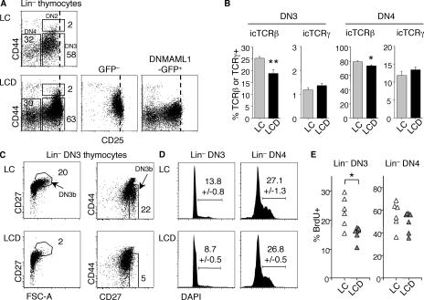
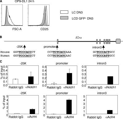
Genetic inactivation of Notch signaling in CD4(-)CD8(-) double-negative (DN) thymocytes was previously shown to impair T cell receptor (TCR) gene rearrangement and to cause a partial block in CD4(+)CD8(+) double-positive (DP) thymocyte development in mice. In contrast, in vitro cultures suggested that Notch was absolutely required for the generation of DP thymocytes independent of pre-TCR expression and activity. To resolve the respective role of Notch and the pre-TCR, we inhibited Notch-mediated transcriptional activation in vivo with a green fluorescent protein-tagged dominant-negative Mastermind-like 1 (DNMAML) that allowed us to track single cells incapable of Notch signaling. DNMAML expression in DN cells led to decreased production of DP thymocytes but only to a modest decrease in intracellular TCRbeta expression. DNMAML attenuated the pre-TCR-associated increase in cell size and CD27 expression. TCRbeta or TCRalphabeta transgenes failed to rescue DNMAML-related defects. Intrathymic injections of DNMAML(-) or DNMAML(+) DN thymocytes revealed a complete DN/DP transition block, with production of DNMAML(+) DP thymocytes only from cells undergoing late Notch inactivation. These findings indicate that the Notch requirement during the beta-selection checkpoint in vivo is absolute and independent of the pre-TCR, and it depends on transcriptional activation by Notch via the CSL/RBP-J-MAML complex.


Figures

Figure 1.
Figure 1.
Characterization of LCD mice. (A) Structure of the ROSA26 DNMAMLf/+ locus. Triangles represent loxP sites. SA, splice acceptor; tpA, trimer of SV40 polyadenylation sequence; bpA, bovine growth hormone polyadenylation sequence. (B) GFP expression in LCD thymocyte subsets. (C) Flow cytometric analysis of LC and LCD thymi. Numbers in B and C represent the percentage of cells in the indicated areas. (D) Absolute number of thymocyte subsets in LC and LCD mice. Numeric data represent means ± SEM (n = 8). *, P < 0.01 according to the Student's t test. γδ T, all CD3+ TCRγ+ T cells (including CD8α+).
Figure 2.
Figure 2.
Lin thymocyte subsets in LCD mice. (A) Flow cytometric analysis of LC/LCD Lin thymocytes. Boxed regions identify CD44+CD25+ DN2, CD44CD25+ DN3, and CD44CD25 DN4 cells. CD25 expression in DN2-DN3 cells was higher in LCD than in LC mice (dashed line). Numbers represent the percentage of cells in the indicated areas. (B) Expression of i.c. TCRβ (LC, n = 11; LCD n = 12) and TCRγ (LC, n = 8; LCD, n = 9) in DN3 and DN4 cells from LC and LCD mice. (C) Decreased DN3b population in LCD versus LC DN3 cells. CD27hiFSChi or CD27hiCD44lo DN3 cells have been defined as DN3b thymocytes (reference 15). Plots are gated on total (GFP and GFP+) Lin DN3 cells. (D) Cell cycle analysis of LC/LCD Lin DN3 and DN4 cells (n = 7 and 8, respectively). Numbers in C and D represent the percentage of cells in the indicated areas. (E) BrdU incorporation after a 3-h labeling (n = 6 and 7, respectively). Numeric data represent means ± SEM. *, P < 0.05; or **, P < 0.01 according to the Student's t test.
Figure 3.
Figure 3.
Loss of canonical Notch signaling results in decreased cell size and CD25 expression. (A) Sorted LC or LCD GFP+ DN3 thymocytes were cultured with OP9-DL1 cells expressing the Notch ligand Delta-like 1. Flow cytometric analysis was performed after 24 h. (B) Structure of the Il2ra(Cd25) locus showing three conserved putative CSL/RBP-J binding sites. (C) ChIP assay from purified DN3 cells using anti-Notch1 (αNotch1), antiacetylated histone 4 (αAcH4), or control rabbit antibodies, followed by quantitative PCR. Input DNA (aliquot of preimmunoprecipitation–sheared chromatin) was used for normalization. Numeric data represent means ± SEM.
Figure 4.
Figure 4.
Tcrb and Tcra/b transgenes do not rescue DN to DP differentiation in the absence of Notch signaling in vivo. LC and LCD were bred to Tcrb or Tcra/b (TcrAND) tg mice. F1 progeny were compared with parental tg mice. (A) Total thymocyte numbers in Tcrb tg (n = 7), Tcrb tg x LCD (n = 7), and LCD mice (n = 8). Numeric data represent means ± SEM. (B) CD4/CD8 expression in Tcrb tg, Tcrb tg x LCD, and LCD thymi (top) and GFP expression in DP cells (bottom). Numbers represent the percentage of cells in the indicated areas. (C) Total thymocyte numbers in TcrAND tg and TcrAND tg x LCD mice (n = 5). Numeric data represent means ± SEM. (D) CD4/CD8 expression in TcrAND tg and TcrAND tg x LCD thymi. Numbers represent the percentage of cells in the indicated areas.
Figure 5.
Figure 5.
Intrathymic injections reveal an absolute requirement for Notch signaling during β-selection in vivo. (A) Lin CD25+CD44 DN3 cells from donor CD45.2+ LC or LCD mice were injected intrathymically into sublethally irradiated B6-CD45.1+ mice. (left) Percentage of donor-derived CD45.2+ cells after 10 d. (middle) CD4/CD8 expression on donor-derived cells. (right) GFP expression on donor-derived DP cells. A representative example is shown (LC DN3, n = 5; LCD GFP+ DN3, n = 8; LCD GFP DN3, n = 3). Numbers represent the percentage of cells in the indicated areas. (B) Percentage of donor-derived DP cells for each recipient. **, P < 0.05 according to the Student's t test.

References

    1. Radtke, F., A. Wilson, S.J. Mancini, and H.R. MacDonald. 2004. Notch regulation of lymphocyte development and function. Nat. Immunol. 5:247–253. - PubMed
    1. Maillard, I., T. Fang, and W.S. Pear. 2005. Regulation of lymphoid development, differentiation and function by the Notch pathway. Annu. Rev. Immunol. 23:945–974. - PubMed
    1. Nam, Y., P. Sliz, L. Song, J.C. Aster, and S.C. Blacklow. 2006. Structural basis for cooperativity in recruitment of MAML coactivators to Notch transcription complexes. Cell. 124:973–983. - PubMed
    1. Radtke, F., A. Wilson, G. Stark, M. Bauer, J. van Meerwijk, H.R. MacDonald, and M. Aguet. 1999. Deficient T cell fate specification in mice with an induced inactivation of Notch1. Immunity. 10:547–558. - PubMed
    1. Sambandam, A., I. Maillard, V.P. Zediak, L. Xu, R. Gerstein, J. Aster, W.S. Pear, and A. Bhandoola. 2005. Notch signaling controls the generation and differentiation of early T lineage progenitors. Nat. Immunol. 6:663–670. - PubMed

Publication types

MeSH terms