

Inflammation in carotid atherosclerotic plaque: a dynamic contrast-enhanced MR imaging study
- PMID: 16966482
- PMCID: PMC1820770
- DOI: 10.1148/radiol.2412051336
Inflammation in carotid atherosclerotic plaque: a dynamic contrast-enhanced MR imaging study
Abstract
Purpose: To prospectively evaluate if there is an association between plaque enhancement at magnetic resonance (MR) imaging and proinflammatory cardiovascular risk factors and plaque content.
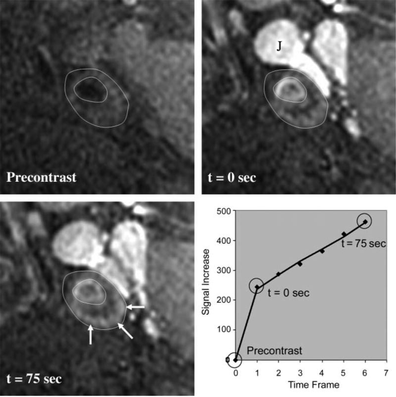
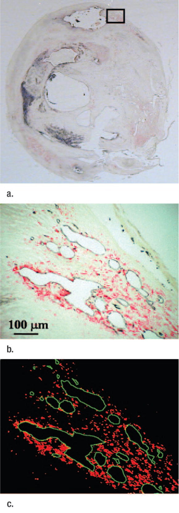
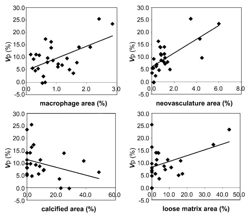
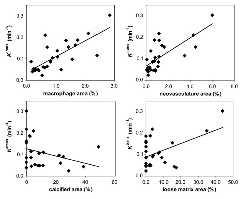
Materials and methods: This study was performed with informed consent, HIPAA compliance, and institutional review board approval. Contrast agent dynamics within carotid plaques were measured in 30 patients (29 men, one woman; mean age, 67.7 years +/- 10.7 [standard deviation]) who were scheduled to undergo carotid endarterectomy. Measurements were based on kinetic modeling of images obtained at 15-second intervals during which a gadolinium-based contrast agent was injected. The time-varying signal intensities within the plaques were used to estimate the fractional plasma volume (vp) and transfer constant (Ktrans) of contrast material into the extracellular space. Pearson correlation coefficients were computed between blinded MR measurements and histologic measurements of plaque composition, including macrophages, neovasculature, necrotic core, calcification, loose matrix, and dense fibrous tissue. Correlation coefficients or mean differences were computed regarding clinical markers of cardiovascular risk.
Results: Analyzable MR images and histologic results were obtained in 27 patients. Measurements of Ktrans correlated with macrophage (r = 0.75, P < .001), neovasculature (r = 0.71, P < .001), and loose matrix (r = 0.50, P = .01) content. Measurements of v(p) correlated with macrophage (r = 0.54, P = .004), neovasculature (r = 0.68, P < .001), and loose matrix (r = 0.42, P = .03) content. For clinical parameters, significant associations were correlated with Ktrans only, with decreased high-density lipoprotein levels (r = -0.66, P < .001) and elevated Ktrans measurements in smokers compared with nonsmokers (mean, 0.134 min(-1) vs 0.074 min(-1), respectively; P = .01).
Conclusion: The correlations between Ktrans and histologic markers of inflammation suggest that Ktrans is a quantitative and noninvasive marker of plaque inflammation, which is further supported by the correlation of Ktrans with proinflammatory cardiovascular risk factors, decreased high-density lipoprotein levels, and smoking.
Figures
References
-
- Kerwin W, Hooker A, Spilker M, et al. Quantitative magnetic resonance imaging analysis of neovasculature volume in carotid atherosclerotic plaque. Circulation. 2003;107:851–856. - PubMed
-
- Yuan C, Kerwin WS, Ferguson MS, et al. Contrast enhanced high resolution MRI for atherosclerotic carotid artery tissue characterization. J Magn Reson Imaging. 2002;15:62–67. - PubMed
-
- Wasserman BA, Smith WI, Trout HH, et al. Carotid artery atherosclerosis: in vivo morphologic characterization with gadolinium-enhanced double-oblique MR imaging—initial results. Radiology. 2002;223:566–573. - PubMed
-
- Wasserman BA, Casal SG, Astor BC, Aletras AH, Arai AE. Wash-in kinetics for gadolinium-enhanced magnetic resonance imaging of carotid atheroma. J Magn Reson Imaging. 2005;21:91–95. - PubMed
-
- Weiss CR, Arai AE, Bui MN, et al. Arterial wall MRI characteristics are associated with elevated serum markers of inflammation in humans. J Magn Reson Imaging. 2001;14:698–704. - PubMed
Publication types
MeSH terms
Substances
Grants and funding
LinkOut - more resources
Full Text Sources
Other Literature Sources
Medical
Miscellaneous
