. 2006 Oct;149(4):355-64.
doi: 10.1038/sj.bjp.0706883. Epub 2006 Sep 11.

Sustained contraction and loss of NO production in TGFbeta1-treated endothelial cells

Affiliations

Sustained contraction and loss of NO production in TGFbeta1-treated endothelial cells

M Watanabe et al. Br J Pharmacol. 2006 Oct.

Abstract

Background and purpose: Transforming growth factor beta1 (TGFbeta1) is generated in atherosclerotic and injured vessel walls. We examined whether the endothelial-to-mesenchymal transdifferentiation induced by TGFbeta1 affects endothelial functions.

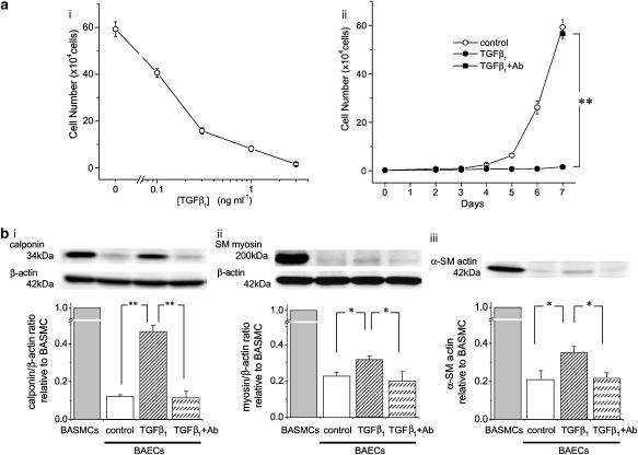
Experimental approach: Bovine aortic endothelial cells (BAECs) were treated with 3 ng ml(-1) TGFbeta1 for 7 days. Contraction of TGFbeta1-treated BAECs was assessed by collagen gel contraction assay. Protein expression and phosphorylation were assessed by Western blotting. Intracellular Ca2+ concentration and NO production were measured using fura2 and DAF-2, respectively.

Key results: TGFbeta1-treated BAECs showed dense actin fibers and expressed smooth muscle marker proteins; they also changed into smooth muscle-like, spindle-shaped cells in collagen gel cultures. ATP (10 microM) induced a gradual contraction of collagen gels containing TGFbeta1-treated BAECs but not of gels containing control BAECs. ATP-induced contraction of TGFbeta1-treated BAECs was not reversed by the removal of ATP but was partially suppressed by a high concentration of sodium nitroprusside (1 microM). TGFbeta1-treated BAECs showed sustained phosphorylation of myosin light chain in response to ATP and low levels of basal MYPT1 expression. ATP-induced Ca2+ transients as well as eNOS protein expression were not affected by TGFbeta1 in BAECs. However, ATP-induced NO production was significantly reduced in TGFbeta1-treated BAECs. Anti-TGFbeta1 antibody abolished all of these TGFbeta1-induced changes in BAECs.

Conclusions and implications: Mesenchymal transdifferentiation induced by TGFbeta1 leads to sustained contraction and reduced NO production in endothelial cells. Such effects, therefore, would not be beneficial for vascular integrity.


Figures

Figure 1
Figure 1
TGFβ1-induced growth inhibition and expression of smooth muscle markers in BAECs. (a) A fixed number of cells (2000 cells) were seeded at day 0, and the cell numbers were manually counted after culturing for 7 days in the absence or presence of various concentrations of TGFβ1 (i), or for 2–7 days in the absence or presence of 3 ng ml−1 TGFβ1 (ii). In some experiments, anti-TGFβ1 antibody (300 ng ml−1) was pre-incubated with 3 ng ml−1 TGFβ1 for 1 h at room temperature and applied to the cells (ii, closed square). **P<0.01. n=5–7. (b) Western blotting of the expressions of calponin (i), SM myosin (ii) and α-SM actin (iii) in BASMCs and control, TGFβ1 (3 ng ml−1)-treated and TGFβ1/antibody-treated BAECs. Expression of β-actin was also measured as an internal control. The cells were cultured in each condition for 7 days. Representative band images are shown in the upper panels. Densitometric analysis of smooth muscle markers/β-actin ratios relative to BASMCs are shown in the lower panels (n=4). *P<0.05, **P<0.01.
Figure 2
Figure 2
Time-dependent change in actin fibres in BAECs. Control culture medium (a) or culture medium containing TGFβ1 (3 ng ml−1, b) or TGFβ1/antibody (c) was applied to subconfluent BAECs. Cytosolic actin cytoskeleton was then stained with rhodamine-phalloidin after 1 h, 24 h or 7 days. Higher magnification images of ‘7 day' cells are taken from the dotted areas of the corresponding lower magnification images. Actin fibres in BASMCs are also shown (d). Scale bar in each panel indicates 50 μm.
Figure 3
Figure 3
Gel contraction assay. BAECs were cultured on a plate for 7 days in the absence or presence of 3 ng ml−1 TGFβ1, and harvested by trypsinization. Cells were then embedded in collagen gels that were overlaid with the same culture media. BASMCs were also embedded in collagen gels. Cell images in the gels after 3 days are shown in (a). Scales, 50 μm. ATP (10 μM)-induced contractions of these gels were assessed by measuring the surface area of the gels (b, n=6–8). See Materials and methods for a detailed protocol.
Figure 4
Figure 4
Effects of SNP and ET-1 on gel contraction. (a) Gels containing BASMCs or TGFβ1-treated BAECs were contracted with 10 μM ATP, and the effects of sequential application of 10 nM and 1 μM SNP were examined (closed symbols, n=4–6). Control data were obtained without SNP application from the same gel preparation (open symbols, n=4–6). Note that 10 nM SNP induced relaxation only in gels containing BASMCs. (b) ET-1 (1 nM) induced contraction in gels with BASMCs, but not in gels containing control or TGFβ1-treated BAECs (n=4–6).
Figure 5
Figure 5
Effects of ATP on p-MLC in BAECs and BASMCs. BAECs were cultured on a plate with control medium or medium containing TGFβ1 (3 ng ml−1) or TGFβ1/antibody for 7days. BASMCs were also cultured for 7days. Total cellular proteins were collected before (0 min) or during 10 μM ATP application and after removing ATP at the times indicated. Expression levels of p-MLC and total MLC were then assessed with Western blotting. The upper panels show the representative band images of p-MLC, and the lower panel shows the densitometric analysis of p-MLC/total MLC values relative to the 0 min value (n=5).
Figure 6
Figure 6
Western blot analysis of the expressions of MYPT1 and p-MYPT1 in BAECs and BASMCs. Total cellular proteins were prepared as described in the legend to Figure 5. Basal expression levels of MYPT1 and β-actin before ATP application are shown in (a) band images show data from a representative experiment and the bars show the densitometric analysis of MYPT1 expression relative to β-actin from six experiments (*P<0.05, **P<0.01). Expression of p-MYPT1 before, during and after ATP (10 μM) application are shown in (b). Band images show a representative result and the graph shows the time-dependent change in p-MYPT1/total MYPT values, relative to the initial (time=0) value (n=5).
Figure 7
Figure 7
ATP (10 μM)-induced Ca2+ transients in BAECs. [Ca2+]i was measured in control and TGFβ1-treated BAECs. Ca2+ traces from representative cells are shown in (a). The bar graphs in (b) show the statistical analysis of the peak value (i) and time-integral (ii) of the ATP-induced net [Ca2+]i increment. Numbers in parenthesis indicate the number of measurements. ns, P>0.05.
Figure 8
Figure 8
NO production in control and TGFβ1-treated BAECs. (a) Expression of eNOS and β-actin proteins were analysed with Western blotting in control and TGFβ1-treated BAECs. Band images show representative data and the bar graph shows the mean eNOS/β-actin values, relative to day 0 control (n=5). ns, P>0.05. (b) ATP (10 μM)-induced NO production was assessed with DAF-2 fluorescence. Cells were excited at 490 nm wavelength every 30 s, and fluorescence intensity at 515 nm was measured. DAF-2 fluorescence relative to its initial value (time 0) was then averaged. Open and closed circles show the data from control (n=22) and TGFβ1-treated (n=20) BAECs, respectively. Anti-TGFβ1 antibody restored NO production (closed square, n=18). **P<0.01, TGFβ1 vs TGFβ1/antibody.

Similar articles

Cited by

References

    1. Arciniegas E, Neves CY, Carrillo LM, Zambrano EA, Ramirez R. Endothelial-mesenchymal transition occurs during embryonic pulmonary artery development. Endothelium. 2005;12:193–200. - PubMed
    1. Arciniegas E, Sutton AB, Allen TD, Schor AM. Transforming growth factor β1 promotes the differentiation of endothelial cells into smooth muscle-like cells in vitro. J Cell Sci. 1992;103:521–529. - PubMed
    1. Assoian RK, Fleurdelys BE, Stevenson HC, Miller PJ, Madtes DK, Raines EW, et al. Expression and secretion of type β transforming growth factor by activated human macrophages. Proc Natl Acad Sci USA. 1987;84:6020–6024. - PMC - PubMed
    1. Ehrlich HP, Rockwell WB, Cornwell TL, Rajaratnam JB. Demonstration of a direct role for myosin light chain kinase in fibroblast-populated collagen lattice contraction. J Cell Physiol. 1991;146:1–7. - PubMed
    1. Emmert DA, Fee JA, Goeckeler ZM, Grojean JM, Wakatsuki T, Elson EL, et al. Rho-kinase-mediated Ca2+-independent contraction in rat embryo fibroblasts. Am J Physiol. 2004;286:C8–C21. - PMC - PubMed

Publication types

MeSH terms