

Computer tomography assessment of pedicle screw insertion in percutaneous posterior transpedicular stabilization
- PMID: 16967297
- PMCID: PMC2213548
- DOI: 10.1007/s00586-006-0221-x
Computer tomography assessment of pedicle screw insertion in percutaneous posterior transpedicular stabilization
Abstract
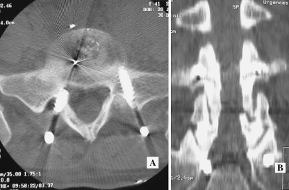
Percutaneous insertion of cannulated pedicle screws has been recently developed as a minimally invasive alternative to the open technique during instrumented fusion procedures. Given the reported rate of screw misplacement using open techniques (up to 40%), we considered it important to analyze possible side effects of this new technique. Placement of 60 pedicle screws in 15 consecutive patients undergoing lumbar or lumbosacral fusion, mainly for spondylolisthesis, were analyzed. Axial, coronal, and sagittal reformatted computer tomography images were examined by three observers. Individual and consensus interpretation was obtained for each screw position. Along with frank penetration, we also looked at cortical encroachment of the pedicular wall by the screw. Thirteen percent of the patients (2/15) had severe frank penetration from the screws, while 80% of them (12/15) had some perforation. On axial images the incidence of severe frank pedicle penetration was 3.3% while the overall rate of screw perforation was 23%. In coronal images the overall screw perforation rate rose to 30% while the rate of severe frank pedicle penetration remained unchanged. One patient (6.6%) suffered S1 root symptoms due to a frankly medially misplaced screw, requiring re-operation. This study has shown that percutaneous insertion of cannulated pedicle screws in the lumbar spine is an acceptable procedure. The overall rate of perforation in axial images is below the higher rates reported in the literature but does remain important. Frank penetration of the pedicle was nevertheless low. It remains a demanding technique and has to be performed with extreme care to detail.
Figures

References
-
- Foley KT, Gupta SK. Percutaneous pedicle screw fixation of the lumbar spine: preliminary clinical results. J Neurosurg. 2002;97:7–12. - PubMed
Publication types
MeSH terms
LinkOut - more resources
Full Text Sources
Medical
