. 2006 Oct;66(12):1365-77.
doi: 10.1002/neu.20315.

Tissue inhibitor of metalloproteinase-2 (TIMP-2) regulates neuromuscular junction development via a beta1 integrin-mediated mechanism

Affiliations

Tissue inhibitor of metalloproteinase-2 (TIMP-2) regulates neuromuscular junction development via a beta1 integrin-mediated mechanism

Gentian Lluri et al. J Neurobiol. 2006 Oct.

Abstract

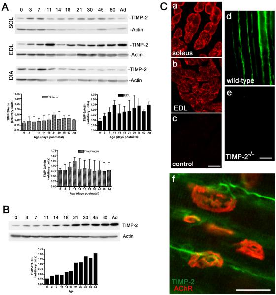
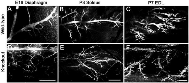
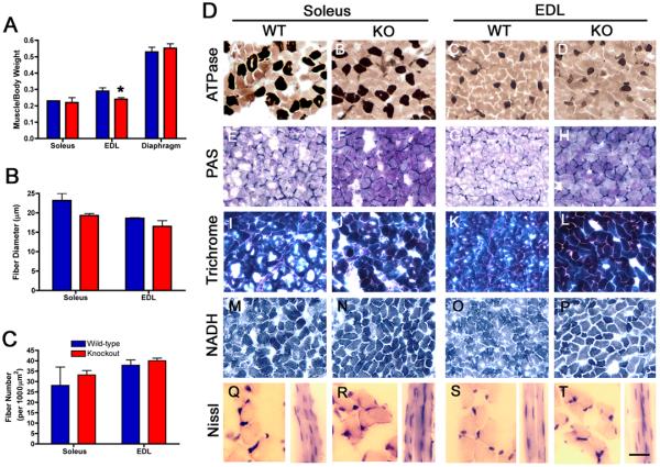
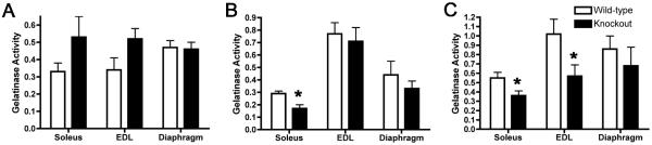
Extracellular matrix (ECM) molecules play critical roles in muscle function by participating in neuromuscular junction (NMJ) development and the establishment of stable, cytoskeleton-associated adhesions required for muscle contraction. Matrix metalloproteinases (MMPs) are neutral endopeptidases that degrade all ECM components. While the role of MMPs and their inhibitors, the tissue inhibitor of metalloproteinases (TIMPs), has been investigated in many tissues, little is known about their role in muscle development and mature function. TIMP-2 -/- mice display signs of muscle weakness. Here, we report that TIMP-2 is expressed at the NMJ and its expression is greater in fast-twitch (extensor digitorum longus, EDL) than slow-twitch (soleus) muscle. EDL muscle mass is reduced in TIMP-2-/- mice without a concomitant change in fiber diameter or number. The TIMP-2-/- phenotype is not likely due to increased ECM proteolysis because net MMP activity is actually reduced in TIMP-2-/- muscle. Most strikingly, TIMP-2 colocalizes with beta1 integrin at costameres in the wild-type EDL and beta1 integrin expression is significantly reduced in TIMP-2-/- EDL. We propose that reduced beta1 integrin in fast-twitch muscle may be associated with destabilized ECM-cytoskeletal interactions required for muscle contraction in TIMP-2-/- muscle; thus, explaining the muscle weakness. Given that fast-twitch fibers are lost in muscular dystrophies and age-related sarcopenia, if TIMP-2 regulates mechanotransduction in an MMP-independent manner it opens new potential therapeutic avenues.


Figures

Figure 1
Figure 1. TIMP-2 is expressed at the neuromuscular junction
A) Western blot analysis of soleus (SOL), extensor digitorum longus (EDL), and diaphragm (DIA) (25 μg crude homogenate) at the indicated postnatal ages (Ad-adult). TIMP-2 expression was normalized to actin as a loading control. TIMP-2 expression is greatest in EDL and is increased during development. B) Western blot analysis of spinal cord lumbar enlargement (20 μg crude homogenate). TIMP-2 is more abundantly expressed in spinal cord than muscle. Like the EDL, TIMP-2 expression in spinal cord is developmentally up-regulated. C) Confocal photomicrographs of TIMP-2 expression in transverse sections of P21 soleus (a, c), EDL (b), and longitudinal sections of P14 sartorius (d, e). Antibody specificity is demonstrated by the lack of immunolabeling with secondary antibody alone (c) and in TIMP-2−/− muscle (e). In addition to labeling muscle basal lamina, TIMP-2 (green) labels motor neurons at the NMJ endplate (red, rhodamine-conjugated α- BTX) (f). Scale bars = 25 μm.
Figure 2
Figure 2. Intramuscular nerve branching is increased in TIMP-2−/− mice
Confocal micrographs show neurofilament-145 immunolabeling in TIMP-2−/− mice and wild-type littermate controls. A well-defined central nerve trunk with branches emanating from it is present in wild-type E16 diaphragm (A) and P3 soleus (B), while the P7 EDL shows a discretely organized endplate band (C). This is in stark contrast to the nerve branching in TIMP-2−/− mice. The altered TIMP-2−/− nerve morphology may be due to increased nerve branching (D) and/or axon defasciculation (E); thereby, resulting in a disorganized endplate region (F). Altered nerve branching is also present in the diaphragm at P3, but not P7, and in the P3 EDL and P7 soleus (data not shown). Data are representative of 3 embryos and 6 neonatal mice. Scale bar = 100 μm.
Figure 3
Figure 3. Synapse elimination is unaltered in TIMP-2−/− mice
A) Synapse elimination was determined by quantitating the percent of monoinnervated endplates in the diaphragm. Monoinnervated endplates in wild-type controls and TIMP-2−/− littermates is similar at P3 (45.6 ± 0.5% vs. 43.3 ± 2.0%; p = 0.3, n = 3), P7 (74.4 ± 1.9% vs. 79.1 ± 4.2%; p = 0.3, n = 4), and P14 (93.7 ± 1.9% vs. 94.0 ± 1.3%; p = 0.9, n = 3). B) Confocal photomicrographs of P3 diaphragm demonstrate that the number of axons per polyinnervated endplate is also comparable in wild-type and TIMP-2−/− mice. C) Multiphoton micrograph demonstrating the nerve terminal with FM1-43 (green) and the AChR endplate region with rhodamine-conjugated α-BTX (red) indicates that pre- and post-synaptic NMJ components are appropriately aligned in the diaphragm of P21 wild-type and TIMP-2−/− mice. Scale bars = 50 μm (B), 25 μm (C).
Figure 4
Figure 4. Muscle cytoarchitecture is normal in P21 TIMP-2−/− muscle
A) Muscle weight, normalized to body weight, is reduced in TIMP-2−/− EDL (* p = 0.02), but not soleus (p = 0.5) or diaphragm (p = 0.5, n = 5). B) Fiber diameter is not reduced in soleus (wt: 21.4 ± 1.8, ko: 19.2 ± 0.5, p = 0.2) or EDL (wt: 18.4 ± 0.3, ko: 17.4 ± 1.2, p = 0.5, n = 3). C) The number of muscle fibers (per 10,000 μm2) is also not different in the soleus (wt: 28.2 ± 8.9, ko: 33.2 ± 2.2; p = 0.6) or EDL (wt: 37.9 ± 2.5, ko: 40 ± 1.3; p = 0.5). D) TIMP-2−/− muscles at P21 appear histologically normal; with the exception of increased basement membrane glycoproteins and collagen detected with Periodic Acid Schiff (PAS, E-H) and Masson Trichrome stain (I-L), respectively. ATPase at pH 4.3 shows no changes in slow-twitch muscle fiber number or distribution (A-D). In addition, no difference in NADH diaphorase is present (M-P). Examination of muscle cross sections reveals that TIMP-2−/− muscle lack central nuclei, but longitudinal sections appear to possess more nuclei per muscle fiber (Q-T). Scale bar = 50 μm (A-P), 25 μm (Q-T longitudinal sections), and 12.5 μm (Q-T cross sections).
Figure 5
Figure 5. MMP activity increases as wild-type muscles age and is reduced in TIMP-2−/− muscle
MMP activity was determined in P3 (A), P21 (B), and adult (C) muscle. A) At P3, there is a slight increase in MMP activity in TIMP-2−/− soleus and EDL, but this increase did not reach statistical significance (soleus: p = 0.14, EDL: p = 0.07, diaphragm: p = 0.8; n = 5). B) At P21, MMP activity is decreased in the soleus (* p = 0.02) and unchanged in the EDL (p = 0.6) and diaphragm (p = 0.4; n = 4). C) In the adult, MMP activity is decreased in both the soleus (* p = 0.04) and EDL (* p = 0.04), but not diaphragm (p = 0.5; n = 6).
Figure 6
Figure 6. β1 integrin expression is reduced in TIMP-2−/− fast-twitch muscle
A) Immunohistochemistry was performed on perfusion fixed, longitudinal cryosections. TIMP-2 co-localizes with β1 integrin at costameres in P21 EDL (A-C) and to a lesser extent adult EDL (G-I). In the soleus, TIMP-2 and β1 integrin are both expressed in the basal lamina. In addition, TIMP-2 is abundantly expressed in capillary endothelial cells at P21 (D-F) and in the adult (J-L). Scale bar = 25 μm. B) Western blot analysis with 25 μg protein from wild-type and TIMP-2−/− EDL. Densitometry was performed and the relative expression of β1 integrin was normalized to α-tubulin (n=3). The developmental decline in β1 integrin expression that normally occurs in wild-type muscle is much more precipitous in TIMP-2−/− muscle. C) Immunohistochemistry confirms the western blot analysis. β1 integrin expression at P21 is significantly decreased in the EDL and only moderately decreased in the soleus (n=3). Scale bar = 50 μm.

Similar articles

Cited by

References

    1. Bewick GS, Reid B, Jawaid S, Hatcher T, Shanley L. Postnatal emergence of mature release properties in terminals of rat fast- and slow-twitch muscles. Eur. J. Neurosci. 2004;19:2967–2976. - PubMed
    1. Boppart MD, Burkin DJ, Kaufman SJ. α7β1 Integrin Regulates Mechanotransduction and Prevents Skeletal Muscle Injury. Am. J. Physiol. Cell Physiol. 2006 E-pub. - PubMed
    1. Bradford MM. A rapid and sensitive method for the quantitation of microgram quantities of protein utilizing the principle of protein-dye binding. Anal. Biochem. 1976;72:248–254. - PubMed
    1. Brandon EP, Lin W, D’Amour KA, Pizzo DP, Dominguez B, Sugiura Y, Thode S, Ko CP, Thal LJ, Gage FH, Lee KF. Aberrant patterning of neuromuscular synapses in choline acetyltransferase-deficient mice. J. Neurosci. 2003;23:539–549. - PMC - PubMed
    1. Brooks PC, Stromblad S, Sanders LC, von Schalscha TL, Aimes RT, Stetler-Stevenson WG, Quigley JP, Cheresh DA. Localization of matrix metalloproteinase MMP-2 to the surface of invasive cells by interaction with integrin αvβ3. Cell. 1996;85:683–693. - PubMed

Publication types

MeSH terms

Substances