Review
. 2007:47:469-512.
doi: 10.1146/annurev.pharmtox.47.120505.105225.

Diseases caused by defects in the visual cycle: retinoids as potential therapeutic agents

Affiliations
Review

Diseases caused by defects in the visual cycle: retinoids as potential therapeutic agents

Gabriel H Travis et al. Annu Rev Pharmacol Toxicol. 2007.

Abstract

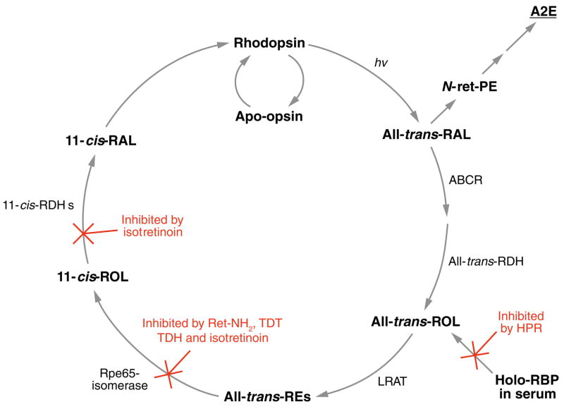
Absorption of a photon by an opsin pigment causes isomerization of the chromophore from 11-cis-retinaldehyde to all-trans-retinaldehyde. Regeneration of visual chromophore following light exposure is dependent on an enzyme pathway called the retinoid or visual cycle. Our understanding of this pathway has been greatly facilitated by the identification of disease-causing mutations in the genes coding for visual cycle enzymes. Defects in nearly every step of this pathway are responsible for human-inherited retinal dystrophies. These retinal dystrophies can be divided into two etiologic groups. One involves the impaired synthesis of visual chromophore. The second involves accumulation of cytotoxic products derived from all-trans-retinaldehyde. Gene therapy has been successfully used in animal models of these diseases to rescue the function of enzymes involved in chromophore regeneration, restoring vision. Dystrophies resulting from impaired chromophore synthesis can also be treated by supplementation with a chromophore analog. Dystrophies resulting from the accumulation of toxic pigments can be treated pharmacologically by inhibiting the visual cycle, or limiting the supply of vitamin A to the eyes. Recent progress in both areas provides hope that multiple inherited retinal diseases will soon be treated by pharmaceutical intervention.


Figures

Figure 1
Figure 1
Structures of visual retinoids (a), N-retinylidene-N-retinyl ethanolamine (A2E) (b), and inhibitors of A2E biogenesis (c).
Figure 2
Figure 2
Visual cycle in retinal pigment epithelium (RPE) cells. Absorption of a photon (hv) by a rhodopsin pigment molecule induces isomerization of 11-cis-RAL to all-trans-RAL resulting in activated metarhodopsin II (MII). Decay of MII yields apo-opsin and free all-trans-RAL. Translocation of all-trans-RAL from the disc membrane to the cytoplasmic space is facilitated by an ATP-binding cassette transporter called ABCR or ABCA4. Mutations in the human ABCA4 gene cause recessive Stargardt macular degeneration. The all-trans-RAL is reduced to all-trans-retinol (all-trans-ROL) by two or more all-trans-ROL dehydrogenases (all-trans-RDHs). Mutations in the gene for one (RDH12) cause Leber congenital amaurosis (LCA) (85). The all-trans-ROL is released by photoreceptors and taken up by RPE cells where it is esterified to a fatty acid by lecithin:retinol acyl transferase (LRAT). Mutations in the LRAT gene cause a severe form of recessive retinitis pigmentosa (RP) (86). The isomerase, RPE-specific 65 kDa protein (Rpe65) uses an all-trans-retinyl ester as substrate to form 11-cis-retinol (11-cis-ROL) plus a free fatty acid. Mutations in the RPE65 gene also cause LCA (87, 88). The Rpe65 isomerase activity may be regulated by the non-photoreceptor opsin, RGR (131). Mutations in the RPE-retinal G protein-coupled receptor (RGR) gene also cause recessive RP (93). 11-cis-ROL binds to the cellular retinaldehyde-binding protein (CRALBP). Mutations in the gene for CRALBP (RLBP1) cause still another form of recessive RP (137). 11-cis-ROL may be esterified by LRAT to yield 11-cis-retinyl esters, a stable storage-form of pre-isomerized retinoid. 11-cis-retinyl esters are hydrolyzed to 11-cis-ROL for synthesis of chromophore by 11-cis-retinyl ester hydrolase (11-cis-REH), which has not yet been cloned. 11-cis-ROL is oxidized to 11-cis-RAL by several 11-cis-ROL dehydrogenases (11-cis-RDHs), including RDH5 and RDH11. Mutations in the gene coding for RDH5 cause delayed dark adaptation and a mild retinal dystrophy called fundus albipunctatus in humans (141). 11-cis-RAL is also bound to CRALBP before being released by the RPE and taken up by the photoreceptor where it recombines with opsin to form rhodopsin.
Figure 3
Figure 3
Hypothesized alternate visual cycle for regeneration of cone pigments. Absorption of a photon (hv) induces 11-cis to all-trans isomerization of the retinaldehyde chromophore in a cone opsin pigment. After dissociation from apo-cone-opsin, the resulting all-trans-RAL is reduced to all-trans-ROL by the NADP+/NADPH-dependent all-trans-ROL dehydrogenase (all-trans-RDH) and released from the photoreceptor outer segment (OS), where it is bound to interstitial retinol binding protein (IRBP) (not shown). The all-trans-ROL released by rods and cones is taken up by Muller cells where it is isomerized to 11- cis-ROL by an isomerase, and esterified to form an 11-cis-retinyl ester by acyl-CoA:retinol acyltransferase (ARAT). These enzyme activities are kinetically coupled and named “isomerosynthase” (35, 36). An 11-cis-retinyl ester is hydrolyzed by 11-cis-retinyl ester hydrolase (REH) to yield 11-cis-ROL, which binds to cellular retinaldehyde-binding protein (CRALBP). 11-cis-REH is regulated by the ratio of apo- to holo-CRALBP in the retinal pigment epithelium (RPE) cells (60). A similar regulatory mechanism is suggested here in Muller cells. 11- cis-ROL released by the Muller cell is bound to IRBP (not shown) and taken up by the cone outer-segment, which contains an NADP+/NADPH-dependent 11-cis-ROL dehydrogenase (11-cis-RDH) activity (35). This activity oxidizes 11-cis-ROL to 11-cis-RAL, which combines with apo-cone-opsin to form a new cone visual pigment. The presence of reciprocal NADP+/NADPH-dependent all-trans-ROL and 11-cis-RDHs in cones affords a self-renewing supply of dinucleotide cofactor at all rates of photoisomerization.
Figure 4
Figure 4
Effects of chromophore supplementation and gene therapy on vision in rpe65−/− and lrat−/−knockout mice. (a) Single-cell recording stimulus response families of rpe−/− mice supplemented with 9-cis-RAL compared with rpe+/+ mice. (b) High performance liquid chromatography (HPLC) analysis of retinoids present in the eyes of lrat−/− mice treated with 9-cis-retinyl acetate. The presented results were reproduced from References and .
Figure 5
Figure 5
Simplified visual cycle showing sites of action for the inhibitors of N-retinylidene-N-retinyl ethanolamine (A2E) formation. All-trans-RAL released by photoactivated rhodopsin condenses with phosphatidylethanolamine (PE) in disc membranes to form N-retinylidene-PE (N-ret-PE). Secondary condensation of N-ret-PE with another all-trans-RAL, followed by electrocyclization, hydrolysis of the phosphate ester, and oxidation yields A2E. Pharmacologic interventions that reduce light-dependent formation of all-trans-RAL greatly reduce the probability of this secondary condensation and inhibit formation of A2E. N-(4-hydroxyphenyl)retinamide (HPR) acts to reduce levels of holo-retinol binding protein (RBP) in serum. Rpe65-mediated isomerization can be blocked by retinylamine (Ret-NH2), the farnesyl-containing analogues, TDH and TDT, and isotretinoin. Isotretinoin also inhibits 11-cis-ROL dehydrogenase (11-cis-RDH).

References

LITERATURE CITED

    1. Filipek S, Stenkamp RE, Teller DC, Palczewski K. G protein-coupled receptor rhodopsin: a prospectus. Annu Rev Physiol. 2003;65:851–79. - PMC - PubMed
    1. Okada T, Ernst OP, Palczewski K, Hofmann KP. Activation of rhodopsin: new insights from structural and biochemical studies. Trends Biochem Sci. 2001;26:318–24. - PubMed
    1. Arshavsky VY, Lamb TD, Pugh EN. G proteins and phototransduction. Annu Rev Physiol. 2002;64:153–87. - PubMed
    1. Rando RR, Bangerter FW. The rapid intermembraneous transfer of retinoids. Biochem Biophys Res Commun. 1982;104:430–36. - PubMed
    1. Groenendijk GW, De Grip WJ, Daemen FJ. Rod outer segment membranes: good acceptors for retinoids? Vision Res. 1984;24:1623–27. - PubMed

RELATED REVIEWS

    1. Mark M, Ghyselinck NB, Chambon P. Function of retinoid nuclear receptors: lessons from genetic and pharmacological dissections of the retinoic acid signaling pathway during mouse embryogenesis. Annu Rev Pharmacol Toxicol. 2006;46:451–80. - PubMed
    1. Chytil F. Rough and rocky road to the retinoid revolution. Annu Rev Nutr. 2003;23:1–16. - PubMed
    1. Palczewski K. G protein-coupled receptor rhodopsin. Annu Rev Biochem. 2006;75:743–67. - PMC - PubMed

Publication types