

Hormonal change and cytokine mRNA expression in peripheral blood mononuclear cells during the development of canine autoimmune thyroiditis
- PMID: 16968404
- PMCID: PMC1809736
- DOI: 10.1111/j.1365-2249.2006.03187.x
Hormonal change and cytokine mRNA expression in peripheral blood mononuclear cells during the development of canine autoimmune thyroiditis
Abstract
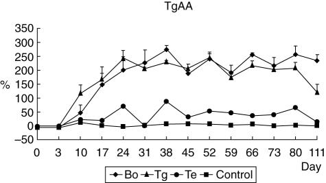
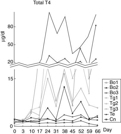
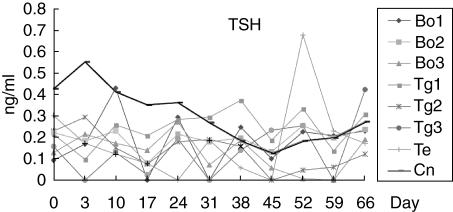
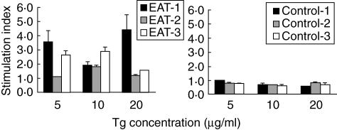
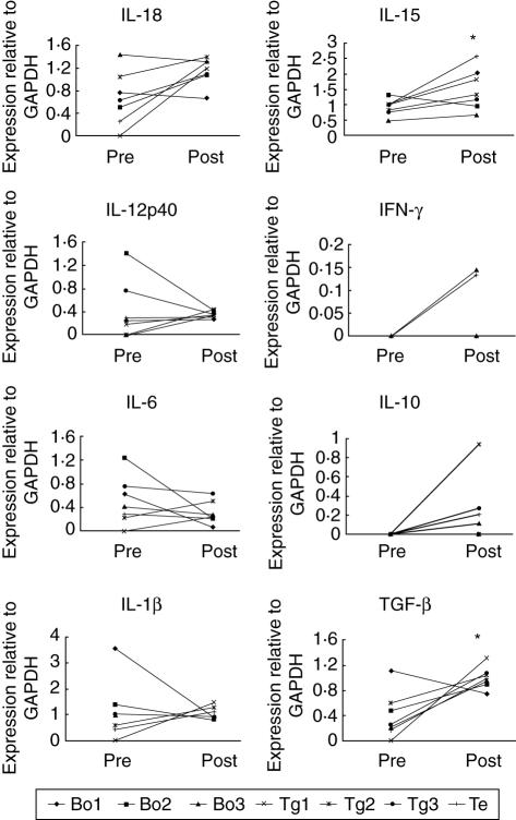
To elucidate the hormonal change and alteration in cytokine expression in peripheral blood mononuclear cells (PBMC) during the early stage of autoimmune thyroiditis, we have developed a canine model of this disease, in which normal dogs were immunized with bovine thyroglobulin (Tg) and/or canine thyroid extract. Serum samples were collected weekly, anti-canine Tg antibody was measured by enzyme-linked immunosorbent assay (ELISA) and thyroid stimulating hormone (TSH) and total T4 levels by radioimmunoassay. We also assayed T lymphocyte proliferation in response to Tg, as well as measuring cytokine mRNA by semiquantitative reverse transcription-polymerase chain reaction (RT-PCR). All six dogs immunized with bovine Tg had both canine Tg autoantibody and anti-T4 antibody. When the sample from the highest TgAA titre time-point was compared with baseline the expression of mRNA encoding the Th1-type cytokine such as interferon (IFN)-gamma, interleukin (IL)-18 and IL-15 was increased during the development of autoimmune thyroiditis. Expression of the Th2-type cytokine, IL-6 showed minimal change and IL-4 expression was not detected in any of the samples. Expression of the T suppressive cytokine, IL-10 and transforming growth factor (TGF)-beta was increased in the presence of antigen stimulation. These findings suggest that, although autoimmune thyroiditis is an organ-specific autoimmune disease, systemic cytokine mRNA expression is also changed.
Figures
Similar articles
-
The effect of gene therapy using CTLA4Ig/silica-nanoparticles on canine experimental autoimmune thyroiditis.J Gene Med. 2008 Jul;10(7):795-804. doi: 10.1002/jgm.1203. J Gene Med. 2008. PMID: 18452240
-
Cytokine gene expression in autoimmune thyroiditis in BioBreeding/Worcester rats.Thyroid. 1999 Oct;9(10):1049-55. doi: 10.1089/thy.1999.9.1049. Thyroid. 1999. PMID: 10560963
-
Induction of oral tolerance in human autoimmune thyroid disease.Thyroid. 1998 Mar;8(3):229-34. doi: 10.1089/thy.1998.8.229. Thyroid. 1998. PMID: 9545109 Clinical Trial.
-
Regulatory B and T cell responses in patients with autoimmune thyroid disease and healthy controls.Dan Med J. 2016 Feb;63(2):B5177. Dan Med J. 2016. PMID: 26836805 Review.
-
Vitiligo and chronic autoimmune thyroiditis.J Med Life. 2021 Mar-Apr;14(2):127-130. doi: 10.25122/jml-2019-0134. J Med Life. 2021. PMID: 34104234 Free PMC article. Review.
Cited by
-
Animal models of Soft Tissue Sarcoma for alternative anticancer therapy studies: characterization of the A-72 Canine Cell Line.Vet Res Commun. 2023 Sep;47(3):1615-1627. doi: 10.1007/s11259-023-10115-z. Epub 2023 Apr 11. Vet Res Commun. 2023. PMID: 37038001 Free PMC article.
-
Co-existence of Hashimoto's thyroiditis with familial Mediterranean fever: is there a pathophysiological association between the two diseases?Clin Exp Immunol. 2009 May;156(2):373-6. doi: 10.1111/j.1365-2249.2009.03891.x. Epub 2009 Feb 19. Clin Exp Immunol. 2009. PMID: 19250274 Free PMC article.
-
Characterization of D-17 Canine Osteosarcoma Cell Line and Evaluation of Its Ability to Response to Infective Stressor Used as Alternative Anticancer Therapy.Animals (Basel). 2020 Oct 28;10(11):1981. doi: 10.3390/ani10111981. Animals (Basel). 2020. PMID: 33126659 Free PMC article.
-
Characterization of MDCK cells and evaluation of their ability to respond to infectious and non-infectious stressors.Cytotechnology. 2020 Feb;72(1):97-109. doi: 10.1007/s10616-019-00360-z. Epub 2019 Dec 4. Cytotechnology. 2020. PMID: 31802289 Free PMC article.
-
Relationships between Cytokine Levels and Disease Parameters during the Development of a Collagen-induced Arthritis Model in Cynomolgus Macaques (Macaca fascicularis).Comp Med. 2019 May 1;69(3):228-239. doi: 10.30802/AALAS-CM-18-000058. Epub 2019 May 8. Comp Med. 2019. PMID: 31068244 Free PMC article.
References
-
- Feldman EC, Nelson RW. Hypothyroidism. In: Feldman EC, Nelson RW, editors. Canine and feline endocrinology and reproduction. 2. Philadelphia: W.B. Saunders Co.; 1996. pp. 187–265.
-
- Catharine RJ, Lynn Guptill-Yoran S. Hypothyroidism. In: Ettinger SJ, Feldman EC, editors. Textbook of veterinary internal medicine. 5. Philadelphia: W.B. Saunders Co.; 2000. pp. 1419–29.
-
- Chistiakov DA, Turakulov RI. CTLA-4 and its role in autoimmune thyroid disease. J Mol Endocrinol. 2003;31:21–36. - PubMed
Publication types
MeSH terms
Substances
LinkOut - more resources
Full Text Sources
Miscellaneous
