. 2006 Nov 10;281(45):34537-48.
doi: 10.1074/jbc.M606654200. Epub 2006 Sep 12.

Control of phospholipid synthesis by phosphorylation of the yeast lipin Pah1p/Smp2p Mg2+-dependent phosphatidate phosphatase

Affiliations

Control of phospholipid synthesis by phosphorylation of the yeast lipin Pah1p/Smp2p Mg2+-dependent phosphatidate phosphatase

Laura O'Hara et al. J Biol Chem. .

Abstract

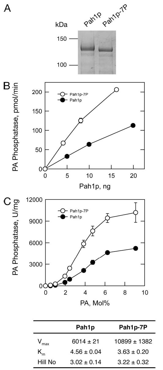
Phosphorylation of the conserved lipin Pah1p/Smp2p in Saccharomyces cerevisiae was previously shown to control transcription of phospholipid biosynthetic genes and nuclear structure by regulating the amount of membrane present at the nuclear envelope (Santos-Rosa, H., Leung, J., Grimsey, N., Peak-Chew, S., and Siniossoglou, S. (2005) EMBO J. 24, 1931-1941). A recent report identified Pah1p as a Mg2+-dependent phosphatidate (PA) phosphatase that regulates de novo lipid synthesis (Han G.-S., Wu, W. I., and Carman, G. M. (2006) J. Biol. Chem. 281, 9210-9218). In this work we use a combination of mass spectrometry and systematic mutagenesis to identify seven Ser/Thr-Pro motifs within Pah1p that are phosphorylated in vivo. We show that phosphorylation on these sites is required for the efficient transcriptional derepression of key enzymes involved in phospholipid biosynthesis. The phosphorylation-deficient Pah1p exhibits higher PA phosphatase-specific activity than the wild-type Pah1p, indicating that phosphorylation of Pah1p controls PA production. Opi1p is a transcriptional repressor of phospholipid biosynthetic genes, responding to PA levels. Genetic analysis suggests that Pah1p regulates transcription of these genes through both Opi1p-dependent and -independent mechanisms. We also provide evidence that derepression of phospholipid biosynthetic genes is not sufficient to induce the nuclear membrane expansion shown in the pah1delta cells.


Figures

FIGURE 1
FIGURE 1. Identification of phosphorylation sites in Pah1p
A, schematic representation of the primary structure of Pah1p. The gray and black boxes indicate the conserved N-lipin and HAD-like domains within Pah1p, respectively. Asterisks denote the Ser/Thr-Pro phospho residues identified in this study. Phosphorylated residues in sequences are in bold. B, sequencing of the phospho-Ser-744/Ser-748 peptide of Pah1p by nanoelectrospray MS/MS. Collision-induced dissociation spectrum of a double-charged ion m/z 838.4 is shown. The spectrum containing a y-ion series (y1-y12) and a b-ion series (b2-b6) confirmed the peptide sequence as QIYLELGSPLASPK. The phosphorylation sites were identified by the 167 mass different between y2-y3, and y6-y7, corresponding to phosphoserine 12 and 8. C, mapping of N-terminal Ser/Thr-Pro phosphorylation sites using the MPM-2 antibody. The indicated Pah1p phosphorylation mutants (asterisks indicate the position of the different Ser/Thr to Ala mutations: S168A/S602A/T723A/S744A/S748A, called Pah1p-5P; S168A/S475A/S602A/T723A/S744A/S748A, called Pah1p-6P; S168A/S475A/T522A/S602A/T723A/S744A/S748A, called Pah1p-7P-C; S110A/S114A/S168A/S602A/T723A/S744A/S748A, called Pah1p-7P; S110A/S114A/S168A/S475A/T522A/S602A/T723A/S744A/S748A, called Pah1p-9P) or the wild-type control, tagged with protein A, were affinity-purified using IgG-Sepharose beads and then digested with tobacco etch virus protease to remove the PtA tag. The various pulldowns were resolved by 10% SDS-PAGE and Western-blotted using the anti-MPM2 antibody. Supernatants from each sample were also analyzed using anti-PtA antibodies. D, the effect of alanine substitutions at the phosphorylation sites on the electrophoretic mobility of Pah1p. Protein extracts from pah1Δ (lane 1) or nem1Δ pah1Δ (lanes 2–9) cells transformed with centromeric plasmids expressing wild type (wt) or Pah1p-PtA mutants as indicated were prepared from logarithmically growing cells and resolved by 7% SDS-PAGE followed by Western blot using anti-PtA antibodies.
FIGURE 2
FIGURE 2. Identification of phosphorylation sites on Pah1p dephosphorylated by the Nem1p-Spo7p complex
A, hyperphosphorylated Pah1p, purified from a culture of pah1Δ nem1Δ spo7Δ expressing Pah1p-PtA by IgG-Sepharose chromatography and tobacco etch virus protease digested to remove the PtA tag was incubated with IgG-Sepharose beads with or without Nem1pPtA-Spo7p complex. Reactions were resolved by 7% SDS-PAGE followed by Coomassie staining, and the Pah1p bands were excised from gels and subjected to liquid chromatography-MS/MS analysis as described under “Experimental Procedures.” B, Pah1p-PtA purified from cells overexpressing the Nem1p-Spo7p complex using the galactose promoter or the control vectors was affinity-purified and resolved by 10% SDS-PAGE followed by Coomassie staining, and the Pah1p-PtA bands were excised from gels and subjected to nanoelectrospray mass spectrometry. In both A and B, the positions of phosphoserine residues identified by mass spectrometry within the two Pah1p samples are highlighted by asterisks (Ser/Thr-Pro sites) or triangles (non-Ser/Thr-Pro sites).
FIGURE 3
FIGURE 3. Phosphorylation of Pah1p on Ser/Thr-Pro sites is required for efficient transcriptional dere-pression of phospholipid biosynthetic genes
A, pah1Δ cells expressing the wild-type PAH1 or the PAH1-7P allele from centromeric vectors were grown in synthetic medium supplemented with 10 Δ g/ml inositol to early logarithmic phase. Cells were then transferred into medium lacking inositol, and the mRNAs of INO1 (left panel), OPI3 (right panel), and RTG2 (control) were quantified at the indicated time points by real time RT-PCR. Values are given as INO1:RTG2 and OPI3:RTG2 ratios. Values and errors were calculated from two independent experiments. WT, wild type. B, serial dilutions of wild-type cells transformed with the indicated plasmids were spotted on synthetic plates lacking inositol and supplemented with glucose (left) or galactose (right). Cells were grown for the indicated times at 30 °C.
FIGURE 4
FIGURE 4. Effect of phosphorylation on Ser/Thr-Pro sites on the PA phos-phatase activity of Pah1p
A, SDS-PAGE of the purified untagged Pah1p and Pah1p-7P from wild-type yeast cells, stained with Coomassie Blue. The positions of the molecular mass standards are indicated. B, the Mg2+-dependent PA phosphatase activity of the purified Pah1p and Pah1p-7P proteins was measured with the indicated protein contents. The values shown were determined from triplicate enzyme determinations (errors fall within the size of the circles). C, effect of PA surface concentration on Pah1p- and Pah1p-7P-dependent PA phosphatase activity. The indicated purified Pah1p and Pah1p-7P proteins from yeast were assayed for Mg2+-dependent PA phos-phatase activity at the indicated surface concentrations (mol %) of PA. The molar concentration was held constant at 0.2 mM. The values shown were determined from triplicate enzyme determinations ± S.D. Lower panel, the kinetic parameters for the Pah1p and Pah1p-7P samples analyzed in B and C are shown.
FIGURE 5
FIGURE 5. Deletion of OPI1 rescues the inositol auxotrophy of GAL-PAH1-7P
Serial dilutions of wild-type (OPI1) or isogenic opi1Δ cells transformed with the indicated plasmids were spotted on synthetic plates lacking inositol and supplemented with glucose (left) or galactose (right). Cells were grown for the indicated times at 30 °C.
FIGURE 6
FIGURE 6. Cooperative effects of pah1Δ and opi1Δ on the derepression of phospholipid biosynthetic genes
A, the mRNA levels of INO1 were analyzed in the wild-type and isogenic pah1Δ, opi1Δ, and pah1Δ opi1Δ strains grown in YEPD medium by quantitative real time RT-PCR. Amplification of each sample was performed in duplicate and normalized to ACT1. Values and errors were calculated from three independent experiments performed on different days. Values are expressed as –fold derepression over the values measured in the wild-type strain. B, the OPI3 transcripts were quantified as in A. C, serial dilutions of the wild-type and isogenic pah1Δ, opi1Δ and pah1Δ opi1Δ strains were spotted on YEPD plates and grown at 30 °C for the indicated times.
FIGURE 7
FIGURE 7. Phospholipid overproduction is not sufficient for nuclear membrane growth
SEC63-GFP and RFP-PUS1 expressed from centromeric plasmids were used to visualize nuclear/ER membrane and nuclear morphology respectively in wild-type cells (A), pah1Δ cells (B), opi1Δ cells (C), wild-type cells transformed with high copy vectors expressing INO2 and INO4 under the control of the inducible MET25 promoter (D) and wild-type cells transformed with a centromeric vector expressing HMG1 under the control of the inducible GAL1/10 promoter (E). Cells in D were grown in medium lacking methionine and in E in medium supplemented with 2% galactose to induce INO2-INO4 and HMG1, respectively. In all cases cells in early logarithmic phase were visualized by confocal microscopy. Bars, 5 μm.

Similar articles

Cited by

References

    1. Nunnari J, Walter P. Cell. 1996;84:389–394. - PubMed
    1. Warren G, Wickner W. Cell. 1996;84:395–400. - PubMed
    1. Hetzer MW, Walther TC, Mattaj IW. Annu Rev Cell Dev Biol. 2005;21:347–380. - PubMed
    1. Prunuske AJ, Ullman KS. Curr Opin Cell Biol. 2006;18:108–116. - PMC - PubMed
    1. Newport JW, Wilson KL, Dunphy WG. J Cell Biol. 1990;111:2247–2259. - PMC - PubMed

Publication types

MeSH terms