. 2006 Sep 19;103(38):14110-5.
doi: 10.1073/pnas.0602587103. Epub 2006 Sep 12.

beta-Arrestin2 mediates nephrin endocytosis and impairs slit diaphragm integrity

Affiliations

beta-Arrestin2 mediates nephrin endocytosis and impairs slit diaphragm integrity

Ivo Quack et al. Proc Natl Acad Sci U S A. .

Abstract

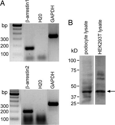
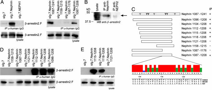
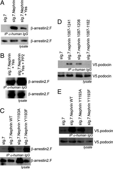
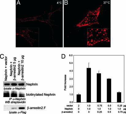
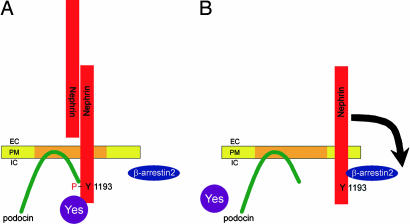
beta-Arrestins mediate internalization of plasma membrane receptors. Nephrin, a structural component of the glomerular slit diaphragm, is a single transmembrane spanning receptor and belongs to the family of adhesion molecules. Its mutation causes a hereditary nephrotic syndrome. We report the previously undescribed interaction of beta-arrestin2 with the nephrin C terminus. The phosphorylation status of nephrin Y1193 regulates inversely the binding of beta-arrestin2 and podocin. The Src-family member Yes, known to enhance podocin-nephrin interaction by nephrin phosphorylation, diminishes beta-arrestin2-nephrin interaction. beta-Arrestin2 induces nephrin endocytosis and attenuates nephrin signaling. This finding suggests that nephrin Y1193 serves as a molecular switch that determines the integrity of the slit diaphragm by functional competition between beta-arrestin2 and podocin. This concept offers a molecular pathomechanism of slit diaphragm distortion and opens therapeutic avenues for glomerular diseases.


Conflict of interest statement

The authors declare no conflict of interest.

Figures

Fig. 1.
Fig. 1.
Podocytes express β-arrestin1 and β-arrestin2. (A) Cultured mouse podocytes show strong RT-PCR signals for β-arrestin1 (169 bp) (Upper) and β-arrestin2 (211 bp) (Lower). (B) In Western blot analysis, endogenous β-arrestin2 can be detected in mouse podocyte lysates and cell lysates of HEK293T cells with β-arrestin2 antiserum at 46 kDa (arrow).
Fig. 2.
Fig. 2.
β-Arrestin2 interacts with the nephrin C terminus. (A) (Left) The C terminus of nephrin but not NEPH1 immobilizes β-arrestin2. HEK293T cells transiently overexpressing heterologous plasma membrane-bound fusion proteins (sIg.7) of the nephrin C terminus, NEPH1, or sIg.7 alone in combination with β-arrestin2 are examined. The coimmunoprecipitated β-arrestin2 is detected by Western blot analysis. To map the interaction site of β-arrestin2 within nephrin, C-terminal truncations are generated. (Right) Only the truncated sIg.nephrin 1087–1208 interacted with β-arrestin2. (B) Endogenous nephrin but not NEPH2 are able to precipitate endogenous β-arrestin2 from mouse kidney lysates. β-Arrestin2 is detected by Western blot analysis with monoclonal β-arrestin2 antisera (arrow). (C) A scheme of the nephrin C-terminal full length with the distribution of tyrosine residues that are predicted to be phosphorylated and the C-terminal nephrin truncations are displayed. Those tyrosine residues that are predicted to be most likely phosphorylated are printed in bold letters [prediction by NetPhos 2.0 (www.cbs.dtu.dk/services/NetPhos)]. The 32 nephrin residues of human, mouse, and rat regulating the β-arrestin2 interaction are aligned (ClustalW). The consensus strength is visualized by colored vertical bars. Tyrosine 1193 (bold) is embedded in a highly cross-species-conserved domain of 8 aa. To map the β-arrestin2 interaction site, the C-terminal truncation sIg.Nephrin 1087–1208 was truncated at the N terminus also. (D) β-Arrestin2 interacts with nephrin through the nephrin amino acids 1112–1120. Amino acids 1087–1111 attenuate the strength of the β-arrestin2–nephrin interaction. (E) Fine mapping delineates the β-arrestin2–nephrin interaction site to the nephrin amino acids 1118–1120 (QWT).
Fig. 3.
Fig. 3.
Nephrin Y1193 is the switch for β-arrestin2 or podocin interaction with nephrin controlled by phosphorylation. (A) Immunoprecipitation of the nephrin C terminus in the presence of Yes decreases the interaction with β-arrestin2. (B) Addition of PP2 reverses the Yes-mediated attenuation of the interaction between nephrin and β-arrestin2. (C) Point mutation of the Y1193 within the nephrin C terminus modulates the interaction with β-arrestin2. The mutation of tyrosine 1193 to alanine (Y1193A) attenuates the interaction with β-arrestin2. The nephrin mutation of tyrosine 1193 to phenylalanine (Y1193F) interacts with β-arrestin2 as strong as nephrin wild type. (D) The podocin interaction with the nephrin C terminus maps to the same nephrin region that regulates the interaction with β-arrestin2. Immunoprecipitation of the nephrin C terminus and its truncations (as labeled) delineates the podocin interaction motif to the nephrin amino acids 1177–1208, the same region that regulates the interaction with β-arrestin2. (E) In contrast to β-arrestin2, podocin interacts with nephrin Y1193A but not with nephrin Y1193F. (Overexpression experiments were done in HEK293T.)
Fig. 4.
Fig. 4.
β-Arrestin2 promotes nephrin endocytosis and regulates nephrin-dependent signaling. (A) Nephrin molecules form numerous small clusters at the cell surface. Nephrin expressed at the surface of vital COS7 cells is labeled with a polyclonal nephrin antiserum followed by anti-rabbit-rhodamine red antisera. The cells are kept at 4°C. (B) After incubation at 37°C for 20 min, the small cell surface nephrin clusters merged to larger intracellular vesicles below the plasma membrane. Images were obtained with confocal microscopy. (C) Coexpression of β-arrestin2 in HEK293T cells diminishes the amount of biotinylated nephrin drastically. HEK293T cells expressing nephrin and β-arrestin2 as labeled were biotinylated. Nephrin was precipitated by nephrin antiserum from whole-cell lysates. The probes were blotted and detected by nephrin, M2-antiserum, or streptavidin, respectively. (D) β-Arrestin2 attenuates nephrin-mediated AP-1 transactivation in a dose-dependent manner. Nephrin-dependent AP-1 transactivation is attenuated by increasing amounts of β-arrestin2. Data sets represent mean values with SD of triplicates that were corrected for transfection efficiency by β-gal activity. AP-1 activation is normalized for the vector control and displayed as fold increase. (Experiments were done in HEK293T.)
Fig. 5.
Fig. 5.
The phosphorylation status of the nephrin Y1193 determines and reflects the slit diaphragm integrity. β-Arrestin2 and podocin interact with nephrin in dependence of the nephrin Y1193 phosphorylation status. (A) Extracellular binding, e.g., homodimerization of nephrin molecules, leads to tyrosine phosphorylation of the nephrin C terminus by Src-kinases (i.e., Yes). Subsequently, podocin interacts with the phosphorylated nephrin, anchors the protein complex in detergent-resistant plasma membranes, and enhances nephrin signaling. (B) As soon as nephrin loses its extracellular-binding partner, it becomes dephosphorylated at nephrin Y1193, terminating the podocin interaction. The nephrin molecule loses its recruitment to detergent-resistant membranes and now interacts with β-arrestin2. This interaction initiates the cell surface disappearance of nephrin and terminates nephrin-mediated signaling. EC, extracellular; PM, plasma membrane; IC, intracellular.

References

    1. Kestila M, Lenkkeri U, Mannikko M, Lamerdin J, McCready P, Putaala H, Ruotsalainen V, Morita T, Nissinen M, Herva R, et al. Mol Cell. 1998;1:575–582. - PubMed
    1. Tryggvason K, Patrakka J, Wartiovaara J. N Engl J Med. 2006;354:1387–1401. - PubMed
    1. Peterson JC, Adler S, Burkart JM, Greene T, Hebert LA, Hunsicker LG, King AJ, Klahr S, Massry SG, Seifter JL. Ann Intern Med. 1995;123:754–762. - PubMed
    1. Shih NY, Li J, Karpitskii V, Nguyen A, Dustin ML, Kanagawa O, Miner JH, Shaw AS. Science. 1999;286:312–315. - PubMed
    1. Boute N, Gribouval O, Roselli S, Benessy F, Lee H, Fuchshuber A, Dahan K, Gubler MC, Niaudet P, Antignac C. Nat Genet. 2000;24:349–354. - PubMed

Publication types

MeSH terms