. 2006 Sep 19;103(38):14170-5.
doi: 10.1073/pnas.0602136103. Epub 2006 Sep 12.

Mechanisms of uropathogenic Escherichia coli persistence and eradication from the urinary tract

Affiliations

Mechanisms of uropathogenic Escherichia coli persistence and eradication from the urinary tract

Indira U Mysorekar et al. Proc Natl Acad Sci U S A. .

Abstract

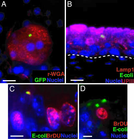
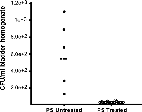
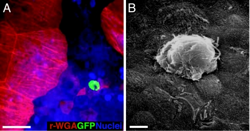
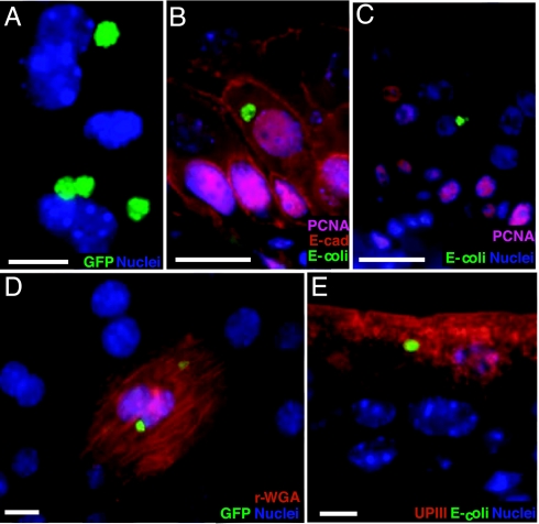
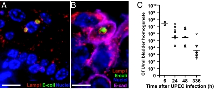
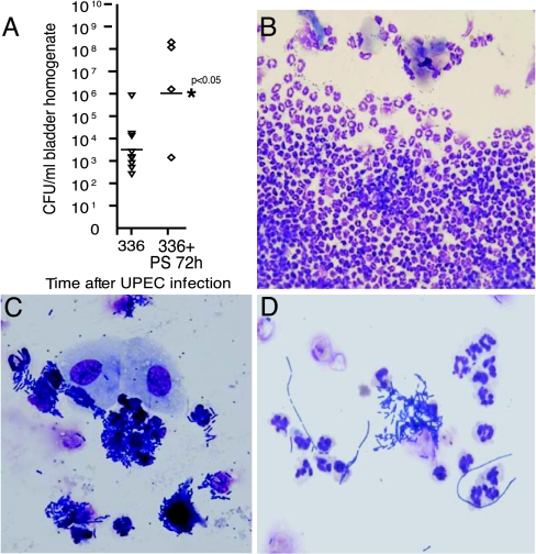
Recurrent urinary tract infections (rUTIs) are a source of considerable morbidity in women. The infecting bacteria in both rUTIs and a de novo acute infection have been thought to originate from an extraurinary location. Here, we show in a murine model of UTI that uropathogenic Escherichia coli (UPEC) established quiescent intracellular reservoirs (QIRs) in Lamp1+ endosomes within the urinary bladder epithelium. Depending on the integrity of the urothelial barriers at the time of initial infection, these QIRs were established within terminally differentiated superficial facet cells and/or underlying transitional epithelial cells. Treatment of infected bladders harboring exclusively superficial facet cell QIRs with the cationic protein, protamine sulfate, led to epithelial exfoliation and eradication of bacteria in 100% of treated animals. However, when the bacterial QIRs were harbored in underlying transitional cells, stimulation of epithelial turnover triggered reemergence of viable organisms and recurrence of infection. Thus, our results suggest (i) that bacterial QIRs within the bladder may be a previously unappreciated source of recurrent UTIs and (ii) that inducing epithelial exfoliation may be a therapeutic avenue for treating this heretofore recalcitrant disease.


Conflict of interest statement

The authors declare no conflict of interest.

Figures

Fig. 1.
Fig. 1.
QIRs form within Lamp1+ vesicles in superficial facet cells. (A) Epifluorescence microscopy on whole-mounted bladders infected with GFP+UTI89 showing bacterial QIRs (green) persisting in differentiated facet cells stained with Alexa Fluor 94-tagged wheat germ agglutinin (r-WGA, to demarcate outlines of facet cell membranes). Epithelial nuclei stained with bis-benzimide appear blue. (Scale bar, 50 μm.) (B) Lamp1+ vesicles (orange, stained with Alexa Fluor 594-tagged rat antibody to Lamp1) containing QIRs (green, stained with Alexa Fluor 88-tagged rabbit polyclonal Abs to E. coli) are in UPIII+ terminally differentiated facet cells (purple with Alexa Fluor 47-tagged mouse mAb to UPIII) 2 weeks after infection. (Scale bar, 50 μm.) The dashed line demarcates epithelial–mesenchymal boundary. (C and D) IFA of tissue sections from BrdU-injected bladders of infected mice show a small BrdU+ (red; stained with Alexa Fluor 94-tagged goat polyclonal antibodies to BrdU) bacterial QIR (yellow) and BrdU+ transitional epithelial cells. A larger BrdU QIR (green) is depicted in D. (Scale bar, 10 μm.)
Fig. 2.
Fig. 2.
Eradication of bacterial QIRs confined to superficial facet cells after PS treatment. CFUs in bladders infected for 2 weeks and treated with 10 mg/ml PS (n = 19 mice) were measured. Control bladders, which were infected but not treated with PS, show colonization (n = 5 mice). Horizontal lines indicate mean titer/mouse/time point.
Fig. 3.
Fig. 3.
UPEC can colonize underlying transitional cells and develop into IBCs. (A) Epifluorescence microscopy on whole-mounted bladders infected with UTI89-GFP reveal an E. coli IBC (green) in underlying transitional cells at 24 hpi. Facet cells are red (r-WGA+). (Scale bar, 50 μm.) (B) Scanning electron micrograph of an IBC at 24 hpi in an underlying transitional cell. (Scale bar, 10 μm.)
Fig. 4.
Fig. 4.
QIRs in underlying transitional cells in PS-pretreated bladders. (A) Whole-mount analysis of PS-pretreated bladders infected for 48 h with UTI89 show GFP+ bacterial QIRs (green). Nuclei appear blue. (B) Bladder tissue sections show that E. coli (green) are intracellular in proliferating PCNA+ (pink nuclei stained with Alexa Fluor 94-linked to goat polyclonal Ab to PCNA); E-cadherin+ underlying cells (red, with Alexa Fluor 94-tagged mouse mAb to E-cadherin, which labels only transitional cells and excludes superficial facet cells). (Scale bar, 10 μm.) (C) UPEC QIRs are also associated with nonproliferating cells (blue nuclei). (D) Whole-mount analysis reveals that the QIRs are GFP+ (green) 2 weeks after infection and can be found in differentiated facet cells (red stain: r-WGA). (E) IFA of bladder tissue sections shows that UPEC QIRs (green) persist in differentiated facet cells (red: UPIII+). (Scale bar, 50 μm in D and E.)
Fig. 5.
Fig. 5.
Subcellular localization of UPEC in Lamp1+ vesicles. (A) Bacterial QIRs (green) are enclosed within Lamp1+ vesicles (red). (B) Lamp1+ vesicles (red) containing UTI89 QIRs (green) are in E-cadherin+ transitional cells (pink) at 48 hpi. (Scale bars, 10 μm.) (C) CFUs in PS-pretreated bladders at various time points after bacterial inoculation. Horizontal lines indicate mean titer/mouse/time point; n = 4–8 mice.
Fig. 6.
Fig. 6.
Reemergence of intracellular UPEC after initiation of epithelial turnover. (A) CFUs in bladders PS-pretreated and UPEC-infected for 2 weeks and subsequently retreated with PS for 72 h. Horizontal lines indicate mean titer/mouse/time point. (B) Urinalysis shows massive neutrophil efflux. (C) Exfoliated urothelial cells and bacteriuria indicating rUTI. (D) Bacterial filaments in rUTI urinalysis.

References

    1. Ronald A. Dis Mon. 2003;49:71–82. - PubMed
    1. Foxman B. Am J Med. 2002;113(Suppl 1A):5S–13S. - PubMed
    1. Foxman B. Am J Public Health. 1990;80:331–333. - PMC - PubMed
    1. Cantey JR. Am J Med. 1985;78:65–75. - PMC - PubMed
    1. Brauner A, Jacobson SH, Kuhn I. Clin Nephrol. 1992;38:318–323. - PubMed

Publication types

MeSH terms

Substances