. 2006 Oct;133(19):3907-17.
doi: 10.1242/dev.02584.

MES-4: an autosome-associated histone methyltransferase that participates in silencing the X chromosomes in the C. elegans germ line

Affiliations

MES-4: an autosome-associated histone methyltransferase that participates in silencing the X chromosomes in the C. elegans germ line

Laurel B Bender et al. Development. 2006 Oct.

Abstract

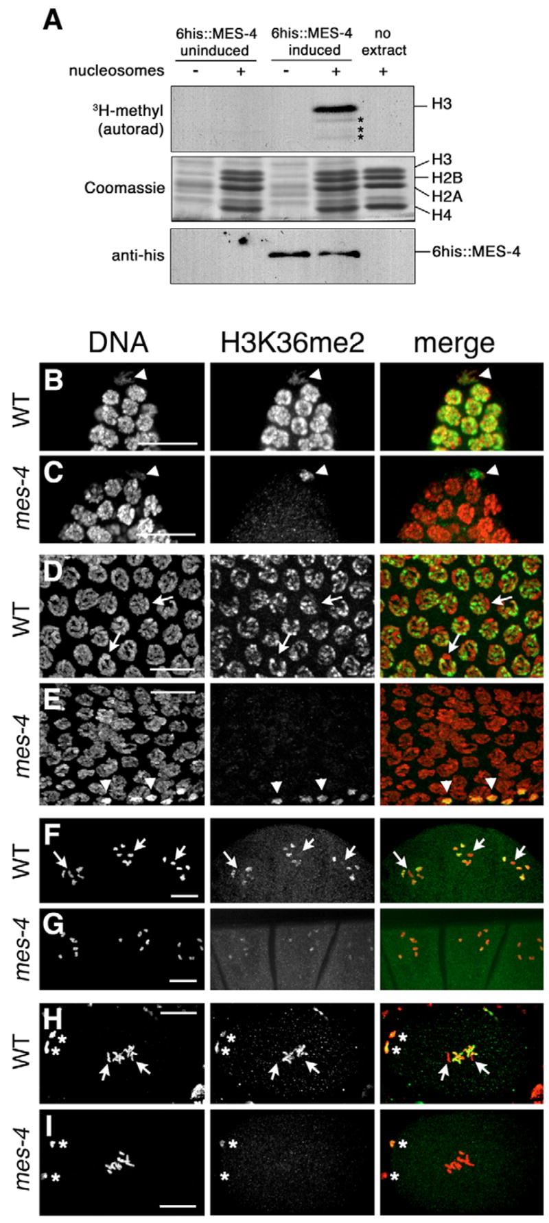
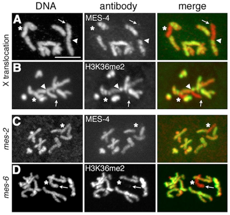
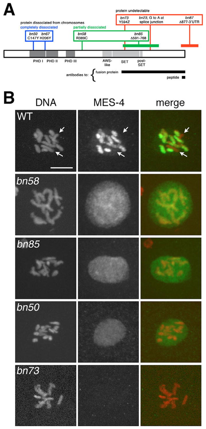
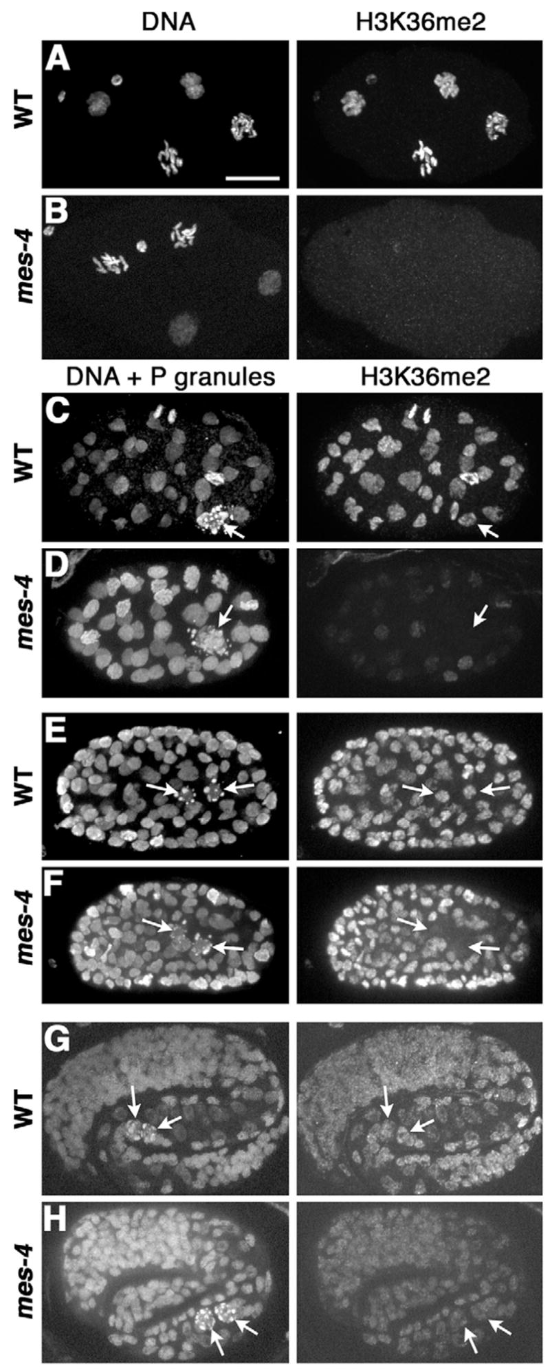
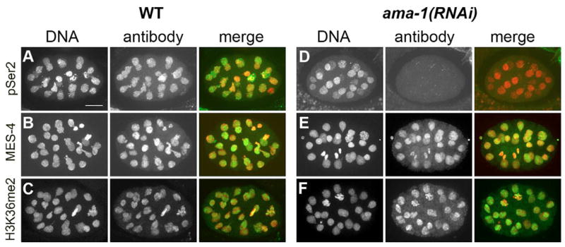
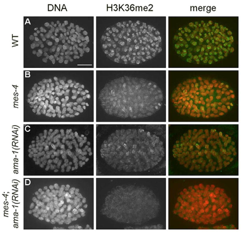
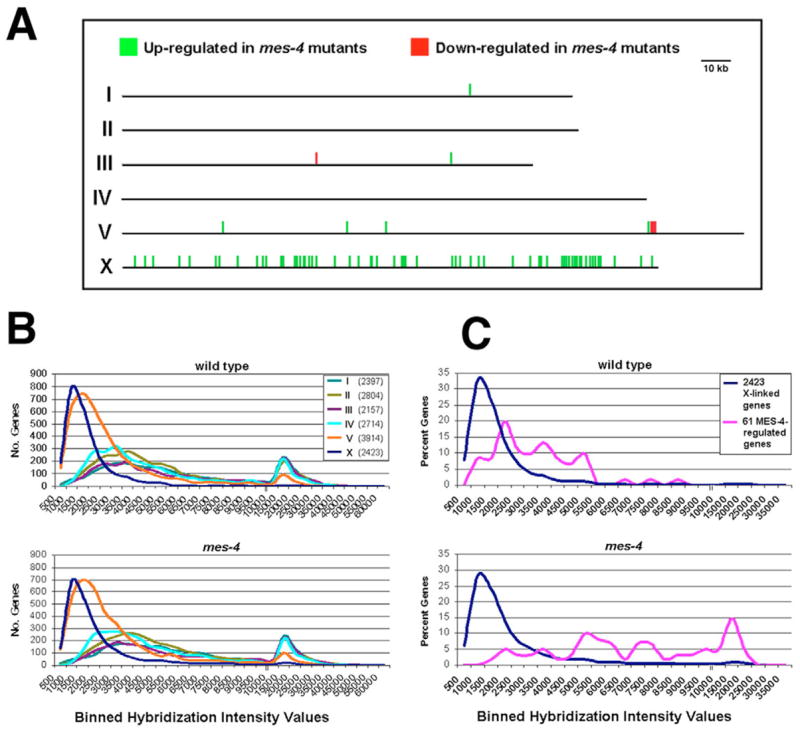
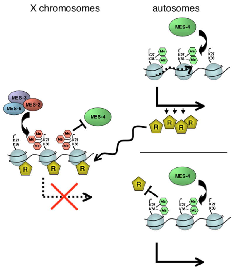
Germ cell development in C. elegans requires that the X chromosomes be globally silenced during mitosis and early meiosis. We previously found that the nuclear proteins MES-2, MES-3, MES-4 and MES-6 regulate the different chromatin states of autosomes versus X chromosomes and are required for germline viability. Strikingly, the SET-domain protein MES-4 is concentrated on autosomes and excluded from the X chromosomes. Here, we show that MES-4 has histone H3 methyltransferase (HMT) activity in vitro, and is required for histone H3K36 dimethylation in mitotic and early meiotic germline nuclei and early embryos. MES-4 appears unlinked to transcription elongation, thus distinguishing it from other known H3K36 HMTs. Based on microarray analysis, loss of MES-4 leads to derepression of X-linked genes in the germ line. We discuss how an autosomally associated HMT may participate in silencing genes on the X chromosome, in coordination with the direct silencing effects of the other MES proteins.


Figures

Fig. 1
Fig. 1. MES-4 is a histone methyltransferase for H3K36
(A) MES-4 has HMT activity in vitro. Bacterially expressed His-tagged MES-4 was incubated with chicken oligonucleosomes and S-adenosyl-L-[methyl-3H] methionine. The reaction products were analyzed by SDS-PAGE followed by fluorography (top panel) and Coomassie staining (middle panel). Western blots (bottom panel) were performed on the soluble extracts to detect MES-4. Asterisks mark H3 breakdown products in lane 4. (B–I) H3K36me2 is undetectable in mes-4(bn73) hermaphrodite germ lines and early embryos. Chromatin (labeled DNA) stained with PA3, red; H3K36me2 staining, green. Arrowheads mark somatic nuclei; arrows indicate X chromosomes. (B,C) Wild-type and mes-4 distal gonads. (D,E) Wild-type and mes-4 pachytene nuclei. (F,G) Wild-type and mes-4 oocytes. (H,I) Wild-type and mes-4 one-cell embryos. Anterior is to the left. Asterisks mark polar bodies. Scale bars: 10 μm.
Fig. 2
Fig. 2. MES-4 and H3K36me2 are concentrated on the autosomes in wild type and spread to the oocyte-derived X chromosome in mes-2 and mes-6 mutant embryos
One-cell embryos with oocyte-derived (anterior, left) and sperm-derived (posterior, right) pronuclei at pronuclear fusion. DNA is stained red with PA3 (A,B,D) or PL4-2 (C); MES-4 or anti-H3K36me2 staining is green. (A,B) Embryos bearing the translocation mnT12 (IV;X) in which the right end of the X chromosome is fused to the left end of chromosome IV. Arrowheads indicate IV;X junctions. Asterisks mark the X portion of the other fused chromosome. (C,D) mes-2(bn11) and mes-6(bn38) embryos. MES-4 and H3K36me2 are on the oocyte-derived X chromosome but are absent from the sperm-derived X (asterisk). Arrows in A,B,D indicate a dot of MES-4 or H3K36me2 at the tip of an X chromosome. Scale bar: 5 μm.
Fig. 3
Fig. 3. mes-4 mutant alleles and the resulting MES-4 distributions
(A) Locations and classifications of the lesions in seven mes-4 alleles. Also see Table S1 in the supplementary material. (B) MES-4 distribution in one nucleus of two-cell wild-type and mes-4 embryos. DNA is stained red with PL4-2 (row 5) or PA3 (all other rows); MES-4 is green. The arrows in the wild-type panels indicate unstained X chromosomes. Scale bar: 5 μm.
Fig. 4
Fig. 4. MES-4 is responsible for all H3K36me2 in early embryos, but another HMT becomes active by the ~40-cell stage
DNA is stained with PL4-2 (G,H) or PA3 (all other rows). P granules are stained with monoclonal antibody OIC1D4 (Hird et al., 1996). Arrows indicate P-granule-containing primordial germ cells. Anterior is to the left. (A,B) Wild-type and mes-4(bn73) four-cell embryos. (C,D) ~40-cell embryos. (E,F) ~100-cell embryos. (G,H) Three-fold embryos. Scale bar: 10μm.
Fig. 5
Fig. 5. MES-4 binding and methylation of H3K36 do not appear to depend on transcription elongation
Comparison of MES-4 and H3K36me2 distribution and levels in ~24-cell wild-type and ama-1(RNAi) embryos. Anterior is to the left. DNA is stained red with anti-acetylated H4 (A,D) or PA3 (B,C,E,F); staining of CTD Ser2 phosphorylation (pSer2), MES-4 and H3K36me2 is green. (A–C) Wild-type embryos. (D) ama-1(RNAi) embryo showing effective depletion of pSer2. (E,F) ama-1(RNAi) embryos showing apparently normal levels and distributions of MES-4 and H3K36me2. Scale bar: 10 μm.
Fig. 6
Fig. 6. Activity of the non-MES-4 H3K36 HMT(s) depends on transcription
Analysis of H3K36me2 levels in ~100-cell embryos lacking MES-4, AMA-1, or both. DNA is stained red with PA3; H3K36me2, green. (A) Wild-type embryo. (B,C) mes-4(bn73) and ama-1(RNAi) embryos display reduced H3K36me2 relative to wild type. (D) The mes-4(bn73); ama-1(RNAi) embryo lacks detectable H3K36me2 signal. Scale bar: 10 μm.
Fig. 7
Fig. 7. Microarray analysis reveals derepression of X-linked genes in mes-4 mutant germ lines
Microarray analysis was performed using linearly amplified RNA from gonads dissected from wild-type and mes-4(bn85) M+Z adult hermaphrodites. (A) Green and red tick marks show the chromosomal positions of the 71 genes that are up- or downregulated at least 1.8-fold in mes-4 mutants relative to wild type (P<0.05, two-tailed paired t-test). Green, 67 genes that are upregulated in mes-4 mutant gonads; red, four genes that are downregulated in mes-4 mutant gonads. Gene IDs and fold differences are shown in Table S2 in the supplementary material. (B) Histograms showing the number of genes on each chromosome that displayed various mean hybridization intensities. The total number of genes sampled on each chromosome is in parentheses in the key. (C) Histograms showing the percentage of X-linked genes that displayed various mean hybridization intensities. Pink, 61 X-linked genes that are upregulated >1.8-fold in mes-4 mutant gonads; blue, all 2423 analyzed genes on the X chromosome. The units on the x-axis change at 10,000.
Fig. 8
Fig. 8. Summary of MES-mediated histone methylation and models for the roles of the MES proteins in silencing the X chromosome in the germ line
(Left) The MES-2/MES-3/MES-6 complex catalyzes di- and trimethylation of H3K27, and preferentially concentrates H3K27me3 on the X chromosome (Bender et al., 2004). We hypothesize that MES-2/MES-3/MES-6 function repels MES-4 from the X chromosome and from regions of the autosomes (Fong et al., 2002) (this study). (Right) Two models for the role of MES-4. (Top right) MES-4 dimethylation of H3K36 in or near an autosomal gene (dotted line) activates that gene to express a repressor (labeled ‘R’) of many X-linked genes. (Lower right) MES-4 concentrates H3K36me2 on the autosomes; MES-4 or H3K36me2, in turn, repels a repressor (labeled ‘R’) from the autosomes, focusing its binding or action on the X chromosome. We speculate that the silencing of genes on the X chromosome is achieved by the combined repressive effects of H3K27me3 and repressor ‘R’ action.

References

    1. Adhvaryu KK, Morris SA, Strahl BD, Selker EU. Methylation of histone H3 lysine 36 is required for normal development in Neurospora crassa. Eukaryot. Cell. 2005;4:1455–1464. - PMC - PubMed
    1. Bannister AJ, Schneider R, Myers FA, Thorne AW, Crane-Robinson C, Kouzarides T. Spatial distribution of di- and tri-methyl lysine 36 of histone H3 at active genes. J Biol Chem. 2005;280:17732–17736. - PubMed
    1. Baugh LR, Hill AA, Slonim DK, Brown EL, Hunter CP. Composition and dynamics of the Caenorhabditis elegans early embryonic transcriptome. Development. 2003;130:889–900. - PubMed
    1. Bean CJ, Schaner CE, Kelly WG. Meiotic pairing and imprinted X chromatin assembly in Caenorhabditis elegans. Nat Genet. 2004;36:100–105. - PMC - PubMed
    1. Bender LB, Cao R, Zhang Y, Strome S. The MES-2/MES-3/MES-6 complex and regulation of histone H3 methylation in C. elegans. Curr Biol. 2004;14:1639–1643. - PubMed

Publication types

MeSH terms