. 2007 Jan 1;109(1):194-202.
doi: 10.1182/blood-2006-02-002873. Epub 2006 Sep 12.

IL-15 and dermal fibroblasts induce proliferation of natural regulatory T cells isolated from human skin

Affiliations

IL-15 and dermal fibroblasts induce proliferation of natural regulatory T cells isolated from human skin

Rachael A Clark et al. Blood. .

Abstract

Regulatory T cells (Tregs) are crucial for the induction and maintenance of self-tolerance and are present in peripheral tissues such as skin and gut under normal, noninflamed conditions. We report isolation and expansion of the Treg population resident in normal human skin. Cutaneous Tregs expressed high levels of CD25, L-selectin, GITR, FOXP3, and intracellular CTLA-4, low levels of CD69, and high levels of the skin-homing addressins CLA, CCR4, and CCR6. Skin Tregs suppressed the proliferation of CD25(lo) T cells from the same skin sample in response to CD3 and CD28 antibodies. Suppression was dependent on cell contact and not affected by neutralizing antibodies to interleukin-10 (IL-10) and transforming growth factor-beta (TGF-beta). Surprisingly, cutaneous Tregs proliferated in an antigen-independent manner when cultured in contact with dermal fibroblasts and IL-15, conditions similar to those found in chronically inflamed skin. We hypothesize that local proliferation of Tregs may occur within inflamed skin and could serve as a brake for cutaneous inflammation as well as a mechanism for the homeostatic proliferation of natural Tregs that has been observed within intact organisms.


Conflict of interest statement

Conflict-of-interest statement: The authors declare they have no conflicts of interest.

Figures

Figure 1
Figure 1
Significant numbers of CD25hiCD69lo skin-resident T cells are isolated from skin explants cultured in IL-2 and IL-15. (A) Skin from a single donor was cultured for 3 weeks on 3D matrices with or without exogenous IL-2 (100 IU/mL) and/or IL-15 (20 ng/mL). Large populations of CD25hiCD69lo T cells were observed in IL-15– and IL-2–treated cultures. (B) Explants cultured in both IL-2 and IL-15 produced the largest absolute number of CD25hiCD69lo T cells. (C) Explants cultured in IL-2 and IL-15 using skin from a second donor; maximal production of CD25hiCD69lo skin-resident T cells occurred at 21 days of culture. Experiments using skin from 5 different donors produced similar results. Numbers indicate the percentage of cells in each quadrant.
Figure 2
Figure 2
CD25hiCD69lo T cells isolated from skin contain a population of natural regulatory T cells. (A) Skin-resident T cells isolated from skin cultured in IL-2 and IL-15 contained increased numbers of FOXP3+ T cells that also expressed high levels of CD25 and low levels of CD69. (B) Skin-resident T cells isolated from explant cultures were sorted into CD25hiCD69lo and CD25lo populations. (C) CD25hiCD69lo T cells (CD25hi) were anergic to stimulation with soluble anti-CD3 and anti-CD28 antibodies (αCD3, CD28) and suppressed the proliferation of CD25lo T cells (CD25lo) isolated from the same sample of skin. (D) Suppression was not affected by neutralizing antibodies to IL-10 (αIL-10) and/or TGF-β (αTGF-β) but was dependent upon cell-cell contact. Suppression was prevented by separation of the CD25hi and CD25lo T-cell populations by a 0.4-μm pore membrane (transwell). (E) A subpopulation of sorted CD25hi T cells retain high expression of CD25 and FOXP3 after 1 week of culture on fibroblast monolayers in the presence of IL-2 and IL-15. Sorted CD25lo cells lack FOXP3+ T cells and FOXP3+ T cells do not develop after 1 week of culture under the same conditions. (F) FOXP3 was up-regulated in both Treg and non-Tregs with cell activation, but this did not obscure identification of Tregs. T cells isolated from skin were examined for FOXP3 expression before and after stimulation with IL-2 and IL-15. Dotplots demonstrate that a clear population of Tregs was discernible under both conditions. The mean fluorescent intensities for each peak are shown on the histograms. FOXP3 expression increased in both groups with stimulation, but the Treg population remained separated from non-Tregs by at least a log increase in FOXP3 staining intensity. For scatterplots, numbers indicate the percentage of cells in each quadrant. For bar graphs, error bars indicate standard deviation.
Figure 3
Figure 3
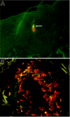
FOXP3+ Tregs are resident in healthy human skin. Sections of healthy human skin were costained with directly conjugated antibodies to CD3 (red) and FOXP3 (green). FOXP3+ Tregs were clearly visible as cells with red membranes and green nuclei. (A) Two T cells present within the epidermis are shown, 1 regulatory (white arrow) and 1 nonregulatory. In general, Tregs were found in the same locations as non-Tregs, located near blood vessels and appendages within the dermis. (B) A particularly brisk infiltrate of T cells containing Tregs surrounding a blood vessel. Such infiltrates can be characteristic of normal human skin.
Figure 4
Figure 4
Phenotype of FOXP3+ natural Tregs isolated from skin. Skin-resident T cells were obtained from skin samples cultured for 3 weeks in IL-2 and IL-15. Unsorted T-cell populations were costained for FOXP3 and the indicated markers. Experiments from 3 different skin donors produced similar results. Numbers indicate the percentage of cells in each quadrant.
Figure 5
Figure 5
IL-15 and dermal fibroblasts induce the proliferation of skin-resident FOXP3+ Tregs. (A) CD25hiCD69lo (CD25hi) skin-resident T cells did not proliferate when treated with IL-2 and IL-15 alone (IL2/15) but proliferated at low levels when stimulated with soluble anti-CD3 and anti-CD28 antibodies (CD3/28). CD25lo skin-resident T cells isolated from the same skin sample also did not proliferate when treated with IL-2 and IL-15 alone, but proliferated robustly after treatment with CD3 and CD28 antibodies. Error bars indicate standard deviation. (B) Sorted CD25hi skin-resident T cells with regulatory activity proliferated when cultured on monolayers of dermal fibroblasts for 1 week in the presence of IL-2 and IL-15. (C) Sorted CFSE-labeled CD25hi skin-resident T cells with regulatory activity cultured on fibroblasts without IL-2 and IL-15 did not proliferate (fib alone) but cells cultured on fibroblast monolayers with IL-2 and IL-15 (fib + IL-2, IL-15) did proliferate. (D) Unsorted CFSE-labeled skin-resident T cells cultured on fibroblast monolayers with IL-2 and IL-15 showed preferential expansion of the CD25hiCD69lo subset. (E) IL-15 and culture on dermal fibroblasts is necessary and sufficient to induce preferential expansion of FOXP3+ skin Tregs. Skin-resident T cells isolated from 2-week explant cultures were labeled with CFSE and cultured for 1 week on fibroblast monolayers with the indicated cytokines. CFSE-low cells have undergone proliferation. All results shown have been replicated using T cells from a minimum of 3 different skin donors. Numbers indicate the percentage of cells in each quadrant.
Figure 6
Figure 6
Skin Treg proliferation requires cell contact with fibroblasts but does not require antigen presentation or costimulation. (A) Skin-resident T cells were cultured in IL-2 and IL-15 for 1 week either in contact with dermal fibroblasts (left panel) or separated from the monolayer by a 0.4 μm transwell membrane (right panel). (B) Dermal fibroblasts do not express HLA-DR, DP, or DQ, nor do they express costimulatory molecules CD80 (B7-1) or CD86 (B7-2). Isotype controls (heavy black line) and test antibodies (▩) histograms are shown. (C) Blockade of HLA-DR, DP, and DQ with neutralizing antibodies did not reduce production of FOXP3+ Tregs from explant cultures. Explant cultures were maintained in IL-2 and IL-15 for 3 weeks; neutralizing antibody was included throughout the culture period and was added with each feeding. Values shown represent the means and SDs of duplicate measurements. Similar results were produced using 2 additional skin donors.
Figure 7
Figure 7
Blood Tregs and non-Tregs both proliferate when cultured with fibroblasts and IL-15. (A) T cells were isolated from the blood, and the highest 5% and lowest 10% of CD25 expressers were isolated by cell sorting and labeled with CFSE. (B) CD25hi T cells contained 58% FOXP3+ Tregs, and a subset of these cells proliferated when cultured for 3 days with IL-15 and fibroblasts. (C) CD25lo T cells contained less than 1% FOXP3+ Tregs; many of these cells proliferated when cultured with IL-15 and dermal fibroblasts. (D) Most CD25hi proliferative cells (P1) were FOXP3+ Tregs, whereas nonproliferative T cells (P0) were purely non-Tregs. (E) Nonregulatory FOXP3 T cells proliferated but did not up-regulate FOXP3 to levels seen in regulatory T cells. For scatterplots, numbers indicate the percentage of cells in each quadrant. (F) Both CLA+ and CLA blood Tregs expand when cultured with fibroblasts, IL-2, and IL-15. T cells were isolated as above, and CD25hi T cells were further separated into CLA+ and CLA subsets. The number of FOXP3+ T cells before and after 1 week of culture with fibroblasts, IL-15, and IL-2 are shown.

Similar articles

Cited by

References

    1. Nishizuka Y, Sakakura T. Thymus and reproduction: sex-linked dysgenesia of the gonad after neonatal thymectomy in mice. Science. 1969;166:753–755. - PubMed
    1. Sakaguchi S, Sakaguchi N, Asano M, Itoh M, Toda M. Immunologic self-tolerance maintained by activated T cells expressing IL-2 receptor alpha-chains (CD25): breakdown of a single mechanism of self-tolerance causes various autoimmune diseases. J Immunol. 1995;155:1151–1164. - PubMed
    1. Hori S, Nomura T, Sakaguchi S. Control of regulatory T cell development by the transcription factor Foxp3. Science. 2003;299:1057–1061. - PubMed
    1. Powell BR, Buist NR, Stenzel P. An X-linked syndrome of diarrhea, polyendocrinopathy, and fatal infection in infancy. J Pediatr. 1982;100:731–737. - PubMed
    1. Satake N, Nakanishi M, Okano M, et al. A Japanese family of X-linked auto-immune enteropathy with haemolytic anaemia and polyendocrinopathy. Eur J Pediatr. 1993;152:313–315. - PubMed

Publication types

MeSH terms

Substances