Review
. 2006 Sep;58(3):281-341.
doi: 10.1124/pr.58.3.3.

International Union of Pharmacology LVIII: update on the P2Y G protein-coupled nucleotide receptors: from molecular mechanisms and pathophysiology to therapy

Affiliations
Review

International Union of Pharmacology LVIII: update on the P2Y G protein-coupled nucleotide receptors: from molecular mechanisms and pathophysiology to therapy

Maria P Abbracchio et al. Pharmacol Rev. 2006 Sep.

Abstract

There have been many advances in our knowledge about different aspects of P2Y receptor signaling since the last review published by our International Union of Pharmacology subcommittee. More receptor subtypes have been cloned and characterized and most orphan receptors de-orphanized, so that it is now possible to provide a basis for a future subdivision of P2Y receptor subtypes. More is known about the functional elements of the P2Y receptor molecules and the signaling pathways involved, including interactions with ion channels. There have been substantial developments in the design of selective agonists and antagonists to some of the P2Y receptor subtypes. There are new findings about the mechanisms underlying nucleotide release and ectoenzymatic nucleotide breakdown. Interactions between P2Y receptors and receptors to other signaling molecules have been explored as well as P2Y-mediated control of gene transcription. The distribution and roles of P2Y receptor subtypes in many different cell types are better understood and P2Y receptor-related compounds are being explored for therapeutic purposes. These and other advances are discussed in the present review.


Figures

Fig. 1
Fig. 1
A phylogenetic tree (dendrogram) showing the relationships among the current members of the P2Y receptor family. The P2Y receptors can be divided into two subgroups, shown with green and blue backgrounds. Sequences were aligned using CLUSTALX, and the tree was built using the TREEVIEW software. Reprinted from Abbracchio et al. (2003) with permission from Elsevier.
Fig. 2
Fig. 2
Conserved residues among P2Y receptors are shown in a larger size font. The residues that have been mutated in the studies of Erb et al. (1995) and Jiang et al. (1997b) are underlined. Those residues that are crucial in the activation of those receptors are in bold. The corresponding sequences of orphan receptors structurally related to P2Y receptors of the second subgroup are also displayed.
Fig. 3
Fig. 3
Structures of adenine-derived nucleotide agonists of P2Y receptors.
Fig. 4
Fig. 4
Structures of nucleotide-based antagonists of P2Y receptors.
Fig. 5
Fig. 5
Structures of non-nucleotide antagonists of P2Y receptors.
Fig. 6
Fig. 6
Structures of uracil-derived nucleotide agonists of P2Y receptors.
Fig. 7
Fig. 7
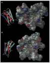
Theoretical structures of the putative nucleotide binding sites of P2Y1 (A) and P2Y12 (B) receptors based on mutagenesis and molecular modeling experiments as described in Costanzi et al. (2004).
Fig. 8
Fig. 8
Examples of analysis of gene activations controlled through P2Y receptor action. A, promoter region of the gene for the catalytic subunit of AChE. The first exon is noncoding, within the 5′-untranslated region. Promoter sites lie on either side of that, illustrated for four of the transcription factors active in muscle cells. Shown are two of the four predicted sites there for the transcription factor Elk-1 (Elk-1 [1] and Elk-1 [3]), which were identified as specific Elk-1 binding sites by gel mobility shift analyses on extracts of myotube nuclei. DNA sequences at those sites were inserted upstream in a luciferase reporter vector for assays as in B. B, two reporter constructs controlled by the specific binding sites for Elk-1 from the AChE gene promoter are used to show that Elk-1 is activated when the P2Y1 or P2Y2 receptors in a muscle cell are stimulated; hence, Elk-1 binds to activate transcription of the AChE gene there. Mouse myotubes transfected with either construct were incubated at 37°C with the agonists shown, or with none (Basal), or with the phorbol ester 12-O-tetradecanoylphorbol-13-acetate (TPA), which produces a nonspecific activation of AChE synthesis in muscle cells. Both Elk-1 sites are seen to have receptor-stimulated promoter activity (and when combined in tandem they are synergistic: not shown). Elk-1 is activated by specific phosphorylation through P2Y1 (2-MeSADP) or P2Y2 (UTP) action. The effect of ATP is significantly higher than effects for the other two (with all three at a saturating concentration), since it acts at both subtypes. C, similar analysis performed using the promoter region of the ε subunit of the nicotinic AChR gene. Both the P2Y1 and P2Y2 receptors act through Elk-1 in activating the multiple genes for the AChR in muscle cells (α, δ, and ε subunit genes have been tested). Note that in confirmation, the P2Y1-specific antagonist MRS2179 blocks this action by 2-MeSADP but not that by UTP (data from Choi et al., 2003; Tung et al., 2004).

References

    1. Abbracchio MP, Boeynaems JM, Barnard EA, Boyer JL, Kennedy C, Miras-Portugal MT, King BF, Gachet C, Jacobson KA, Weisman GA, et al. Characterization of the UDP-glucose receptor (re-named here the P2Y14 receptor) adds diversity to the P2Y receptor family. Trends Pharmacol Sci. 2003;24:52–55. - PMC - PubMed
    1. Abbracchio MP, Burnstock G, Boeynaems J-M, Barnard EA, Boyer JL, Kennedy C, Miras-Portugal MT, King BF, Gachet C, Jacobson KA, et al. The recently deorphanized GPR80 (GPR99) proposed to be the P2Y15 receptor is not a genuine P2Y receptor. Trends Pharmacol Sci. 2005;26:8–9. - PMC - PubMed
    1. Abbracchio MP, Burnstock G. Purinoceptors: are there families of P2X and P2Y purinoceptors? Pharmacol Ther. 1994;64:445–475. - PubMed
    1. Abbracchio MP, Verderio C. Pathophysiological roles of P2 receptors in glial cells. Novartis Found Symp. 2006;276:91–103. - PubMed
    1. Abraham EH, Prat AG, Gerweck L, Seneveratne T, Arceci RJ, Kramer R, Guidotti G, Cantiello HF. The multidrug resistance (mdr1) gene product functions as an ATP channel. Proc Natl Acad Sci USA. 1993;90:312–316. - PMC - PubMed

Publication types

MeSH terms

LinkOut - more resources