. 2006;15(5):369-80.
doi: 10.3727/000000006783981819.

Fate of embryonic stem cells transplanted into the deafened mammalian cochlea

Affiliations

Fate of embryonic stem cells transplanted into the deafened mammalian cochlea

B Coleman et al. Cell Transplant. 2006.

Abstract

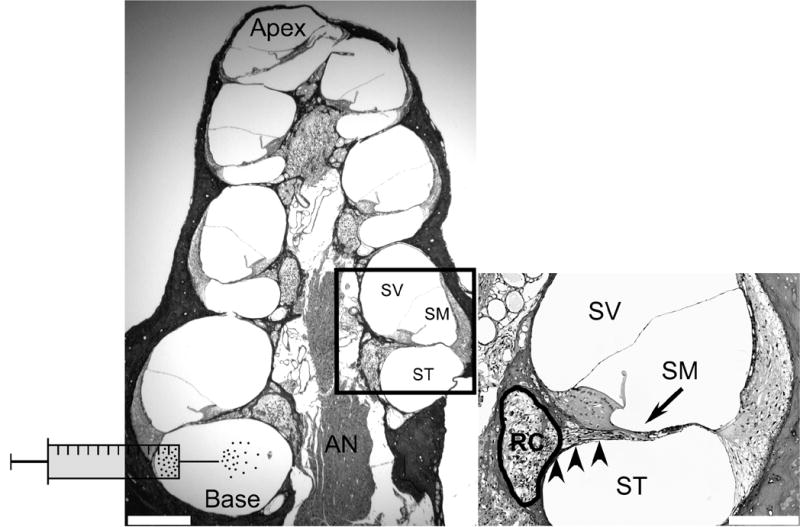
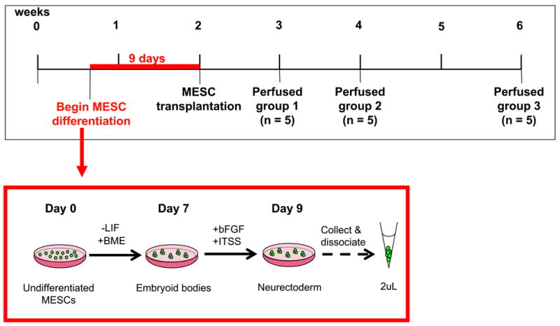
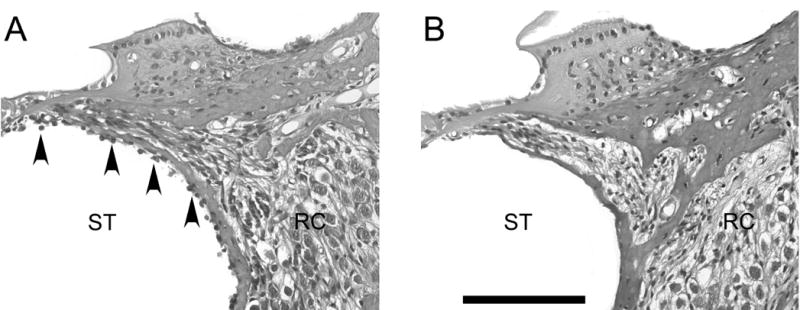
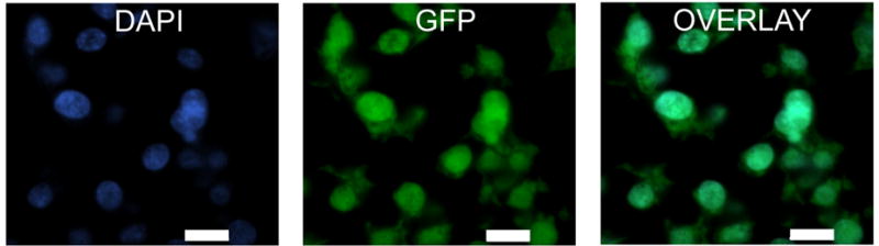
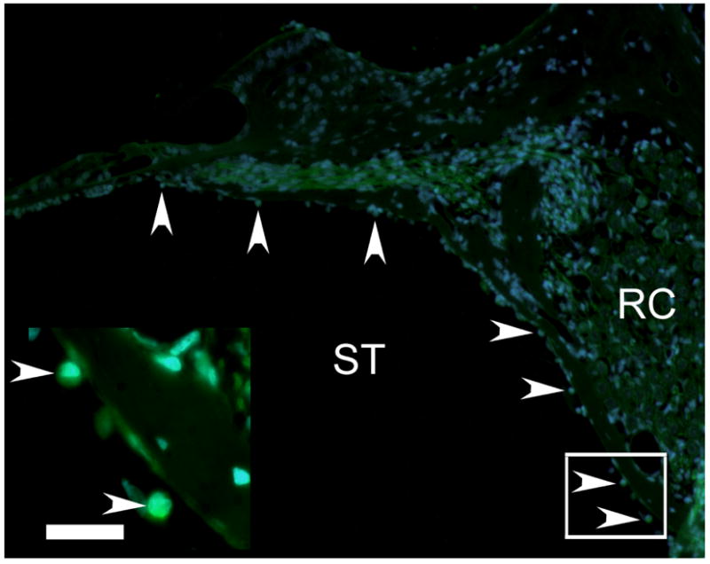
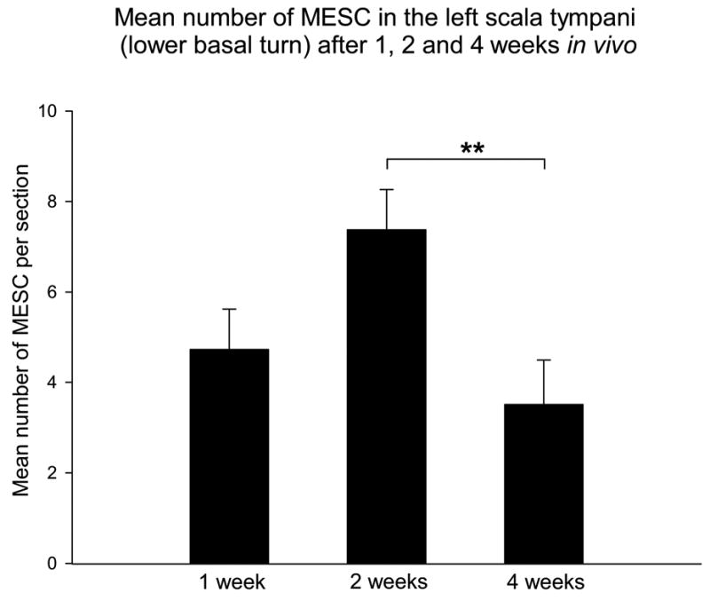
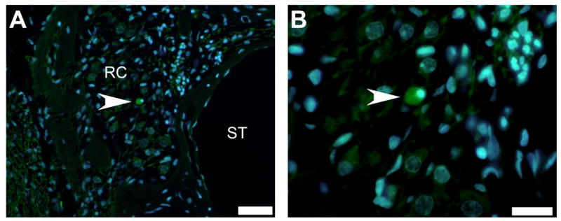
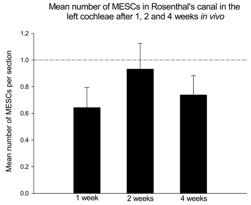
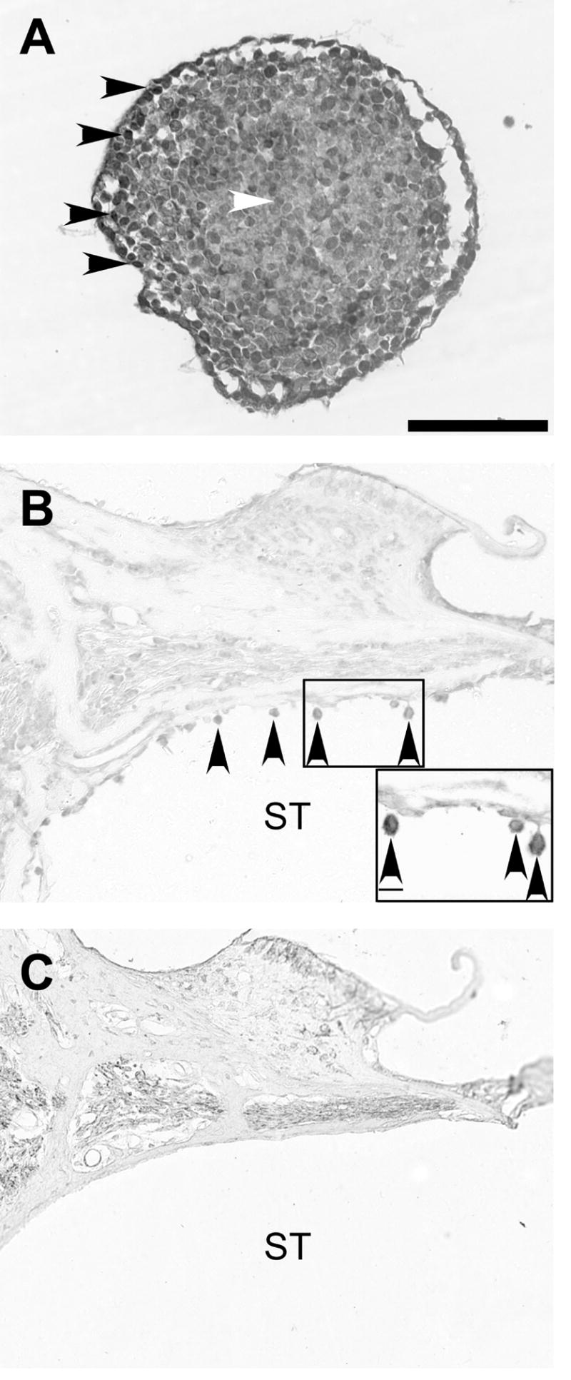
Spiral ganglion neurons (SGNs), the primary afferent neurons of the cochlea, degenerate following a sensorineural hearing loss (SNHL) due to lack of trophic support normally received from hair cells. Cell transplantation is emerging as a potential strategy for inner ear rehabilitation, as injected cells may be able to replace damaged SGNs in the deafened cochlea. An increase in the number of surviving SGNs may result in improved efficacy of cochlear implants (CIs). We examined the survival of partially differentiated mouse embryonic stem cells (MESCs), following xenograft transplantation into the deafened guinea pig cochlea (n=15). Cells were delivered directly into the left scala tympani via microinjection through the round window. Small numbers of MESCs were detected in the scala tympani for up to 4 weeks following transplantation and a proportion of these cells retained expression of neurofilament protein 68 kDa in vivo. While this delivery method requires refinement for effective long-term replacement of damaged SGNs, small numbers of MESCs were capable of survival in the deafened mammalian cochlea for up to 4 weeks, without causing an inflammatory tissue response.


Figures

Figure 1
Figure 1. Anatomy of the deafened guinea pig cochlea and schematic of scala tympani cell delivery technique
Transverse section of a guinea pig cochlea 4 weeks following ototoxic deafening, labelled with H & E. The auditory nerve (AN) is located in the middle of the structure and comprises central processes, which originate from spiral ganglion neurons (SGNs) in Rosenthal’s canal (RC; refer to inset). The AN and RC are collectively referred to as the modiolus. Encircling the modiolus are 4 cochlear turns, each comprising 3 fluid-filled compartments; scala vestibuli (SV), scala media (SM) and scala tympani (ST). The ST and SV are joined at the apex and are filled with the same fluid, perilymph. The SM is filled with endolymph, and is isolated from the ST and SV. MESCs were transplanted into the ST, at the base of the cochlea, as illustrated schematically. Scale bar = 300μm. Inset: Higher magnification photomicrograph illustrating more comprehensively the anatomy of a single cochlear turn. The SV and SM are separated by Reissner’s membrane while the basilar membrane separates the SM and ST. Note the lack of hair cells in deafened animals (arrow), which are normally present at this location on the basilar membrane. SGNs are located within RC (circled) and are degenerating. Peripheral processes extend from SGNs toward the normal site of hair cells (arrow). RC and the ST are separated by a thin bony wall called the osseous spiral lamina (arrowheads). This wall contains numerous bony pores or canaliculae perforantes, which are thought to provide fluid communication channels between the SGNs housed within RC and the ST. Scale bar = 100μm.
Figure 2
Figure 2. Timeline of differentiation and transplantation methodology for MESCs
Twenty guinea pigs were systemically deafened and allowed to recuperate for 2 weeks. Four days after the deafening surgery, undifferentiated MESCs underwent a 9-day induction protocol to sequentially generate embryoid bodies (EBs) and neurectoderm (early neuronal tissue), as described in methods. Two weeks post-deafening, 15 guinea pigs received a 2μL suspension of MESCs, delivered into the left cochlea (Figure 1). A further 5 animals received a transplant of DMEM alone into the left cochlea. The right cochleae of all animals served as a deafened, untreated control. Animals receiving a transplant of MESCs (n=15) were placed randomly into 3 groups (5 animals per group) and perfused 1, 2 or 4 weeks post-stem cell transplantation (group 1, 2 or 3 respectively). Animals receiving a transplant of DMEM (n=5) were perfused 2 weeks post-delivery.
Figure 3
Figure 3. Detection of MESCs in the deafened guinea pig cochlea
Transverse sections of left guinea pig cochleae stained with H & E. MESCs were observed in the scala tympani of transplanted animals for up to 4 weeks post-delivery (arrowheads, A), but not in DMEM treated controls (B). There was no inflammatory tissue response observed in any animals receiving MESC (n=15) or DMEM (n=5) transplants. ST = scala tympani. RC = Rosenthal’s canal. Scale bar = 100μm for both photomicrographs.
Figure 4
Figure 4. Identification of endogenous GFP in MESCs in vitro
High magnification fluorescent photomicrographs showing expression of the nuclear marker DAPI (blue; A), and endogenous GFP (green; B) in MESCs grown in vitro. Co-localisation of DAPI and GFP is illustrated in C. Scale bars = 10μm.
Figure 5
Figure 5. Survival of GFP positive MESCs in vivo
MESCs were detected in the left cochleae of treated animals, using direct fluorescent microscopy for endogenous GFP (green) in combination with the nuclear marker DAPI (blue). Fluorescent photomicrographs illustrate MESCs in the left (treated) cochlea (arrowheads). Inset illustrates higher magnification photomicrograph of the boxed region in the image. ST = scala tympani. RC = Rosenthal’s canal. Scale bar = 20μm.
Figure 6
Figure 6. Mean number of MESCs in the left scala tympani (lower basal turn) after 1, 2 and 4 weeks in vivo
A two-way analysis of variance was used to determine whether there was a statistical difference between the mean number of MESCs in the lower basal turn scala tympani of the left cochleae at 1, 2 and 4 weeks in vivo. This graph illustrates survival of MESCs in the deafened mammalian cochlea for periods up to 4 weeks in vivo, and significantly fewer MESCs in the left cochleae after 4 weeks in vivo. Corresponding p value; ** p=0.01. ST = scala tympani.
Figure 7
Figure 7. Detection of transplanted MESCs within Rosenthal’s canal
A small number of MESCs were detected within Rosenthal’s canal (RC) in the left cochleae (arrowhead, A), using direct fluorescent microscopy for endogenous GFP (green) in combination with the nuclear marker DAPI (blue). Under high magnification these cells were observed to be similar in size and morphology to MESCs in vitro (arrowhead, B). ST = scala tympani. RC = Rosenthal’s canal. Scale bar = 50μm (A) and 20μm (B).
Figure 8
Figure 8. Mean number of MESCs within Rosenthal’s canal after 1, 2 and 4 weeks in vivo
A two-way analysis of variance illustrated there was no significant difference detected between the mean number of MESCs within Rosenthal’s canal in the left cochleae after 1, 2 or 4 weeks in vivo. In addition, the mean number of MESCs detected within Rosenthal’s canal at any time point was never greater than 1 cell per section (perforated line). RC = Rosenthal’s canal.
Figure 9
Figure 9. Neurofilament labeling of MESCs in vitro and in vivo
Light photomicrographs illustrating neurofilament (NF-L) labelling in vitro (A) and in vivo (B, C). NF-L expression in EBs in vitro was strongest around the periphery (black arrowheads, A) and weakest in the centre (white arrowheads, A). Transverse section from the left cochlea 4 weeks following transplantation, demonstrates that a portion of MESCs retained NF-L expression in vivo (arrowheads, B). Neurofilament positive cells were not observed in the scala tympani of DMEM-treated (control) cochleae (C). ST = scala tympani. Scale bar = 100μm (for all photomicrographs). Scale bar for inset (B) = 10μm.

References

    1. Andrew JK. Department of Otolaryngology. Melbourne: University of Melbourne; 2003. Rehabilitation of the Deafened Auditory Nerve With Schwann Cell Transplantation; pp. 1–114.
    1. Coleman B, Hardman J, Crook J, de Silva M, Epp S, Coco A, Shepherd RK. Frontiers in Otorhinolaryngology. Noosa; 2004. Survival and migration of partially differentiated stem cells in the adult guinea pig cochlea.
    1. Coleman B, Hardman J, de Silva M, Crook J, Shepherd RK. International Society for Stem Cell Research. San Francisco; 2005. Strategies for stem cell-based therapy in the mammalian cochlea.
    1. Duckert LG, Duvall AJ., 3rd Cochlear communication routes in the guinea pig-spiral ganglia and osseous spiral laminae: an electron microscope study using microsphere tracers. Otolaryngology. 1978;86:ORL434–446. - PubMed
    1. Ernfors P, Duan ML, ElShamy WM, Canlon B. Protection of auditory neurons from aminoglycoside toxicity by neurotrophin-3. Nature Medicine. 1996;2:463–7. - PubMed

Publication types

Substances